Myofibroblastoma
Key Facts
Terminology
Myofibroblastoma (MFB)
Benign spindle cell mammary tumor, thought to be derived from mammary stromal myofibroblasts
Clinical Issues
Uncommon mammary neoplasm
Typically seen in older patient population
Majority of reported cases of MFB have occurred in male patients; however, overall incidence is likely equal in males and females
Presentation: Painless mass
Complete surgical excision recommended for lesions diagnosed on needle core biopsy
No recurrences have been reported to date
Image Findings
Well-demarcated lobulated mass lesion
Macroscopic Features
Typical lesion about 2 cm in diameter
Microscopic Pathology
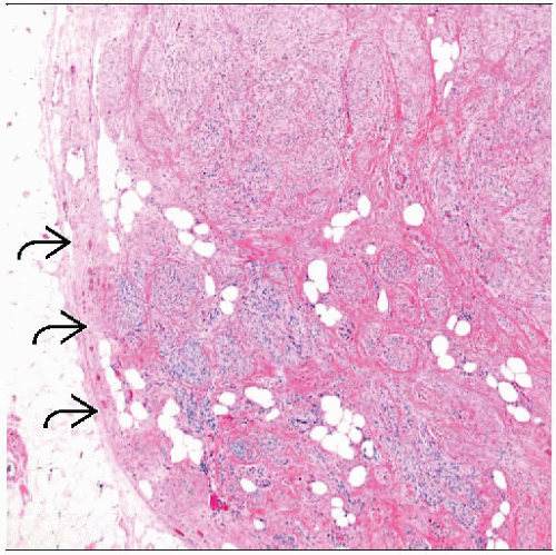
Uniform spindle cell proliferation
Spindle cells closely apposed and arranged in discrete intersecting bundles
Hyalinized bands of collagen separate cells into groups or clusters
Varying degrees of fat cells seen in some lesions
Ancillary Tests
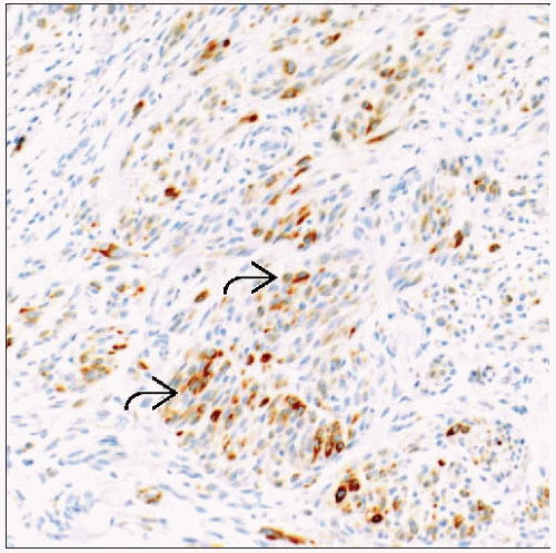
Lesional cells show immunophenotype typical of myofibroblasts
TERMINOLOGY
Abbreviations
Myofibroblastoma (MFB)
Definitions
Benign spindle cell mammary tumor
Original description: “Cells having features of both fibroblasts and smooth muscle”
Derived from mammary myofibroblasts
Cells demonstrate ultrastructural and IHC similarities to myofibroblasts from other sites
Morphologic, structural, and IHC overlap between MFB, spindle cell lipoma, and solitary fibrous tumor of breast
Some authors suggest that these entities represent a spectrum of lesions derived from myofibroblasts
ETIOLOGY/PATHOGENESIS
Hormone Receptors
MFB characteristically express androgen, estrogen, and progesterone receptors
Possible role for sex hormones in pathogenesis of this tumor
MFB may be associated with androgen deprivation in treatment of some prostate cancers
MFB may be associated with gynecomastia or pseudoangiomatous stromal hyperplasia (PASH)
These conditions are also thought to share hormonal etiology
Cytogenetic Abnormalities
Chromosome 13 rearrangements associated with loss of 13q14, partial loss of 16q
These chromosomal alterations are typically also observed in spindle cell lipoma
CLINICAL ISSUES
Epidemiology
Incidence
Uncommon mammary neoplasm; < 1% of mammary tumors
Age
Typically seen in older patient population; peak incidence: 50-75 years
Gender
Initial reports suggested majority of cases of MFB occurred in male patients
Similar lesions have been reported in females
Majority of reported cases of MFB have occurred in male patients; however, overall incidence is likely equal in males and females
Coincidental gynecomastia may be seen in males
Presentation
Palpable painless mass
Mobile
Slow growing
Can be solitary unilateral, bilateral, or multicentric
Lesions with similar appearance can occur in extramammary locations
Treatment
Surgical approaches
Complete surgical excision is recommended for lesions diagnosed on needle core biopsy
Prognosis
Benign stromal tumor
No recurrences have been reported to date in follow-up studies
IMAGE FINDINGS
Ultrasonographic Findings
Circumscribed or lobulated mass
Features similar to those seen in fibroadenoma
MR Findings
Uniform enhancement with internal septation
MACROSCOPIC FEATURES
General Features
Well circumscribed, lobulated, firm or rubbery consistency
Cut surface has pink-tan, homogeneous whorled or nodular appearance
Size
Typical lesion is about 2 cm; most are < 4 cm
MFB up to 10 cm in diameter have been reported
MICROSCOPIC PATHOLOGY
Histologic Features
Uniform spindle cell proliferation
Slender, bipolar or fusiform-shaped spindle cells
Ovoid nuclei, pale cytoplasm
Mitotic figures rare/undetectable
Spindle cells closely apposed and arranged in discrete intersecting bundles and clusters
Well-circumscribed borders
Pushing borders tend to compress normal breast parenchyma at periphery
Circumscribed with no true capsule
Usually no entrapment of ducts&/or lobules
Hyalinized bands of collagen
Distributed throughout lesion
Collagen bands separate spindle cells into groups or clusters
Varying degrees of fat cells seen in some lesions
Rarely, abundant fat suggests lipomatous element or component
Term “lipomatous myofibroblastoma” describes lesions with abundant fatty component
Morphologic and molecular overlap between lipomatous myofibroblastoma and spindle cell lipoma
MFB rarely shows evidence of myoid and cartilaginous differentiation
Rare variant forms of MFB may be encountered
Collagenized or fibrous variant
Epithelioid variant
Can mimic invasive lobular carcinoma, particularly if ER and PR positive on core needle biopsy
Stay updated, free articles. Join our Telegram channel

Full access? Get Clinical Tree


