Mycobacteria


Mycobacteria


Objectives


1. Describe the general characteristics of the Mycobacterium spp., including oxygen requirements, staining patterns and cell morphology, artificial media required for cultivation and growth, and pigmentation.


2. Explain the chemical composition of the bacterial cell wall.


3. Explain the microscopic staining characteristics of Mycobacterium spp. using the Gram stain and acid-fast staining methods.


4. List the most common pathogenic species in the Mycobacterium genus and state the natural habitat, mode of transmission, and reservoir for each.


5. Differentiate M. tuberculosis clinical infections based on the signs and symptoms of the following: primary infection, latent infection, disseminated infection, and reactivation.


6. Compare the current safety and containment methods recommended for handling mycobacterial infectious materials and routine bacteriology in a diagnostic laboratory.


7. Describe the purified protein derivative (PPD; also referred to as the tuberculin skin test). What is the significance of a positive result?


8. List the clinical specimens acceptable for recovery of mycobacteria and describe the limitations of recovery from each type of specimen.


9. Justify the use of DNA probes and molecular sequencing or amplification methods to identify Mycobacterium spp.


10. Evaluate the effectiveness of the staining procedures—Kinyoun, Ziehl-Neelsen, and fluorescent staining (auramine-rhodamine or acridine orange)—for identifying mycobacteria.


11. Describe the requirements for using digestion and decontamination procedures to improve the recovery of Mycobacterium spp.


12. Explain the limitations of digestion and decontamination procedures.


13. Explain the methods commonly used for biochemical identification of Mycobacterium spp. (i.e., niacin, nitrate, urease, modified catalase, Tween 80, tellurite, arylsulfatase, thiophene-2-carboxylic acid hydrazide [TCH], and 5% NaCl tests), including the purpose, principle, and control organisms used for each.


14. Describe the role of the human immunodeficiency virus (HIV) and acquired immunodeficiency syndrome (AIDS) in the dissemination and/or pathogenesis of infections with Mycobacterium spp.


15. Explain the recommended susceptibility testing methods and state when susceptibility testing is required or recommended for Mycobacterium spp.


Traditionally, Mycobacterium spp. have been classified according to phenotypic characteristics. However, since the late 1980s, molecular diagnostics have been used to shift the characterization of these organisms to genotypic studies. This chapter discusses both the phenotypic characterization and the new taxonomy based on molecular genetic data.


The organisms that belong to the genus Mycobacterium are aerobic (although some may grow in reduced oxygen concentrations), non–spore forming (except for M. marinum), nonmotile, very thin, slightly curved or straight rods (0.2 to 0.6 × 1 to 10 µm). Some species may display a branching morphology. Mycobacterium is the only genus in the Mycobacteriaceae family (Actinomycetales order, Actinomycetes class). Genera that are closely related to Mycobacterium include Nocardia, Rhodococcus, Tsukamurella and Gordonia.


Mycobacterium spp. have an unusual cell wall structure. The cell wall contains N-glycolylmuramic acid instead of N-acetylmuramic acid, and it has a very high lipid content, which creates a hydrophobic permeability barrier. Because of this cell wall structure, mycobacteria are difficult to stain with commonly used basic aniline dyes, such as those used in Gram staining. Although these organisms cannot be readily Gram stained, they generally are considered gram positive. However, they resist decolorization with acidified alcohol (3% hydrochloric acid) after prolonged application of a basic fuchsin dye or with heating of this dye after its application. This important property of mycobacteria, which derives from their cell wall structure, is referred to as acid fastness; this characteristic distinguishes mycobacteria from other genera. Rapid-growing mycobacteria (RGMs) may partially or completely lose this characteristic as a result of their growth characteristics.


Another important feature of many species is that they grow more slowly than most other human pathogenic bacteria because of their hydrophobic cell surface. Because of this hydrophobicity, organisms tend to clump, so that nutrients are not easily allowed into the cell. A single cell’s generation time (the time required for a cell to divide into two independent cells) may range from approximately 20 hours to 36 hours for Mycobacterium ulcerans. This slow growth results in the formation of visible colonies in 2 to 60 days at optimum temperature.


Currently, the genus Mycobacterium includes more than 100 recognized or proposed species. These organisms produce a spectrum of infections in humans and animals ranging from localized lesions to disseminated disease. Some species cause only human infections, and others have been isolated from a wide variety of animals. Many species are also found in water and soil.


For the most part, mycobacteria can be divided into two major groups, based on fundamental differences in epidemiology and association with disease: those belonging to the Mycobacterium tuberculosis complex and those referred to as nontuberculous mycobacteria (NTM) (Box 43-1).


Box 43-1   Major Groupings of Organisms Belonging to the Genus Mycobacterium*


Nontuberculous Mycobacteria


*This box is not inclusive; rather, it lists only the prominent mycobacteria isolated from humans.


Mycobacterium Tuberculosis Complex


Tuberculosis was endemic in animals in the Paleolithic period, long before it ever affected humans. This disease (also called consumption) has been known in all ages and climates. For example, tuberculosis was the subject of a hymn in a sacred text from India dating from 2500 BC, and DNA unique to Mycobacterium tuberculosis was identified in lesions from the lung in 1000-year-old human remains found in Peru.


Epidemiology and Pathogenesis


Epidemiology


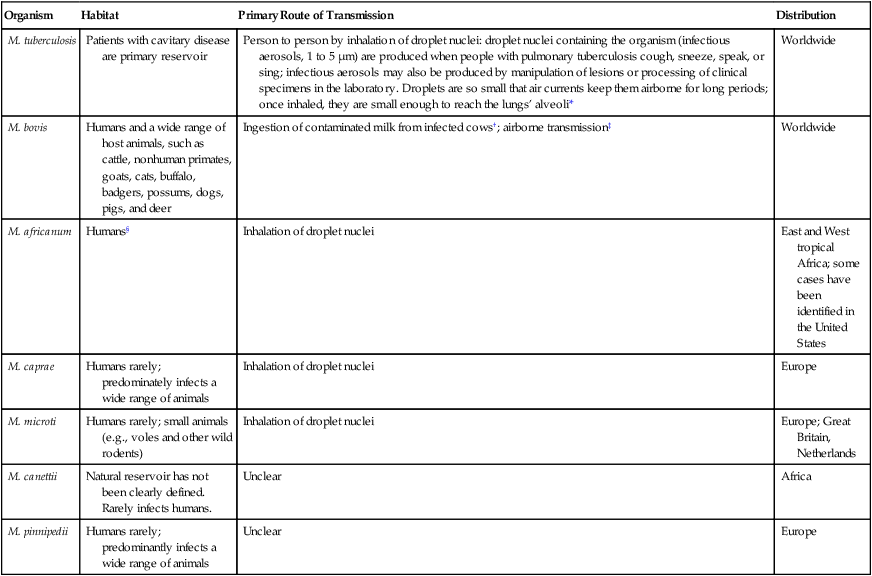
M. tuberculosis is the cause of most cases of human tuberculosis, particularly in developed countries. An estimated 1.7 billion people, or one third of the world’s population, are infected with M. tuberculosis. This reservoir of infected individuals results in 8 million new cases of tuberculosis and 2.9 million deaths annually. Tuberculosis continues to be a public health problem in the United States. An additional complicating factor in the management of tuberculosis is the increasing incidence of co-infection with the human immunodeficiency virus (HIV). HIV-associated tuberculosis remains a significant challenge to world health, with an estimated 1.1 million individuals living with HIV-associated tuberculosis. In the United States, tuberculosis typically is found among the poor, homeless, intravenous (IV) drug users, alcoholics, the elderly, or medically underserved populations. Although the organisms belonging to the M. tuberculosis complex have numerous characteristics in common, including extreme genetic homogeneity, they differ in certain epidemiologic aspects (Table 43-1).


TABLE 43-1


Epidemiology of Organisms Belonging to M. tuberculosis Complex That Cause Human Infections


Organism Habitat Primary Route of Transmission Distribution
M. tuberculosis Patients with cavitary disease are primary reservoir Person to person by inhalation of droplet nuclei: droplet nuclei containing the organism (infectious aerosols, 1 to 5 µm) are produced when people with pulmonary tuberculosis cough, sneeze, speak, or sing; infectious aerosols may also be produced by manipulation of lesions or processing of clinical specimens in the laboratory. Droplets are so small that air currents keep them airborne for long periods; once inhaled, they are small enough to reach the lungs’ alveoli* Worldwide
M. bovis Humans and a wide range of host animals, such as cattle, nonhuman primates, goats, cats, buffalo, badgers, possums, dogs, pigs, and deer Ingestion of contaminated milk from infected cows; airborne transmission Worldwide
M. africanum Humans§ Inhalation of droplet nuclei East and West tropical Africa; some cases have been identified in the United States
M. caprae Humans rarely; predominately infects a wide range of animals Inhalation of droplet nuclei Europe
M. microti Humans rarely; small animals (e.g., voles and other wild rodents) Inhalation of droplet nuclei Europe; Great Britain, Netherlands
M. canettii Natural reservoir has not been clearly defined. Rarely infects humans. Unclear Africa
M. pinnipedii Humans rarely; predominantly infects a wide range of animals Unclear Europe

image


*Infection occasionally can occur through the gastrointestinal tract or skin.


The incidence has decreased significantly in developed countries since the introduction of universal pasteurization of milk and milk products and the institution of effective control programs for cattle.


Can be transmitted human to human, animal to human, and human to animal.


§Infections in animals have not been totally excluded.


Pathogenesis


The pathogenesis of tuberculosis caused by organisms of the M. tuberculosis complex is discussed in Chapter 69. Inhalation of a single viable organism has been shown to lead to infection, although close contact is usually necessary. Of those who become infected with M. tuberculosis, 15% to 20% develop disease. The disease usually occurs some years after the initial infection, when the patient’s immune system breaks down for some reason other than the presence of tuberculosis bacilli in the lung. In a small percentage of infected hosts, the disease becomes systemic, affecting a variety of organs.


After ingestion of milk from infected cows, Mycobacterium bovis may penetrate the gastrointestinal mucosa or invade the lymphatic tissue of the oropharynx. An attenuated strain of M. bovis, bacillus Calmette-Guérin (BCG), has been used extensively in many parts of the world to immunize susceptible individuals against tuberculosis. Because mycobacteria are the classic examples of intracellular pathogens and the body’s response to BCG hinges on cell-mediated immunoreactivity, immunized individuals are expected to react more aggressively against all antigens that elicit cell-mediated immunity. In rare cases, an unfortunate individual’s immune system is so compromised that it cannot handle the BCG, and systemic BCG infection may develop.


Spectrum of Disease


Tuberculosis may mimic other diseases, such as pneumonia, neoplasm, or fungal infections. In addition, clinical manifestations in patients infected with M. tuberculosis complex may range from asymptomatic to acutely symptomatic. Patients who are symptomatic can have systemic symptoms, pulmonary signs and symptoms, signs and symptoms related to other organ involvement (e.g., the kidneys), or a combination of these features. Cases of pulmonary disease caused by M. tuberculosis complex organisms are clinically, radiologically, and pathologically indistinguishable.


Primary tuberculosis typically is considered a disease of the respiratory tract. Common presenting symptoms include low-grade fever, night sweats, fatigue, anorexia (loss of appetite), and weight loss. A patient who presents with pulmonary tuberculosis usually has a productive cough, along with low-grade fever, chills, myalgias (aches), and sweating; however, these signs and symptoms are similar for influenza, acute bronchitis, and pneumonia.


Upon respiratory infection with M. tuberculosis complex organisms, the cellular immune system T cells and macrophages migrate to the lungs, and the organisms are phagocytized by the macrophages. However, these organisms are capable of intracellular multiplication in the macrophages. Often the host is unable to eliminate the organisms, and the result is a systemic hypersensitivity to Mycobacterium antigens. Granulomas or a hard tubercle forms in the lung from the lymphocytes, macrophages, and cellular pathology, including giant cell formation (cellular fusion displaying multiple nuclei). If the Mycobacterium antigen concentration is high, the hypersensitivity reaction may result in tissue necrosis, caused by enzymes released from the macrophages. In this case no granuloma forms, and a solid or semisolid, caseous material is left at the primary lesion site.


In some patients infected with primary active tuberculosis, the disease may spread via the lymph system or hematogenously, leading to meningeal or miliary (disseminated) tuberculosis. This most often occurs in patients with depressed or ineffective cellular immunity.


As previously mentioned, in a small percentage of patients, organs besides the lungs can become involved after infection with M. tuberculosis complex organisms. These organs include the following:


Disseminated tuberculosis may be diagnosed by a positive tuberculin skin test (described later in the chapter).


Patients also may have latent disease (i.e., they have no apparent signs, symptoms, or pathologic condition). A patient with latent tuberculosis is not infectious and does not have active disease, although the organism is present in granulomas. Patients with latent tuberculosis may progress to active disease (also referred to as reactivation of tuberculosis) at any time. Reactivation tuberculosis typically occurs after an incident in which cellular immunity is suppressed or damaged as a result of a change in life style or other health condition.


Individuals infected with HIV are particularly susceptible to developing active tuberculosis. These patients are likely to have rapidly progressive primary disease instead of a subclinical infection.


Diagnosing tuberculosis is more difficult in people infected with HIV, because chest radiographs of the pulmonary disease often lack specificity, and patients frequently are anergic (lack a biologic response) to tuberculin skin testing, a primary means of identifying individuals infected with M. tuberculosis. The tuberculin skin test, or purified protein derivative (PPD) test, is based on the premise that after infection with M. tuberculosis, an individual develops a delayed hypersensitivity cell-mediated immunity to certain antigenic components of the organism. To determine whether a person has been infected with M. tuberculosis, a culture extract of M. tuberculosis (i.e., PPD of tuberculin) is injected intracutaneously. After 48 to 72 hours, an infected individual shows a delayed hypersensitivity reaction to the PPD, characterized by erythema (redness) and, most important, induration (firmness as a result of influx of immune cells). The diameter of induration is measured and then interpreted as to whether the patient has been infected with M. tuberculosis; different interpretative criteria are used for different patient populations (e.g., immunosuppressed individuals, such as those infected with HIV). More recently, the T-Spot TB test (Oxford, Immunotec, United Kingdom) offers next-day results and does not require a follow-up visit with a physician. The assay measures T cells that have been activated by Mycobacterium tuberculosis antigens. Peripheral blood mononuclear cells are incubated with M. tuberculosis-specific antigens stimulating any sensitized T cells in the patient sample. T cell cytokines released in the sample are measured using antibody to capture them and then detected with a secondary antibody conjugated to alkaline phosphatase. This assay should be interpreted in correlation with the patient’s signs and symptoms.


The PPD test is not 100% sensitive or specific, and a positive reaction to the skin test does not necessarily signify the presence of disease. Because of these issues, a new test approved by the U.S. Food and Drug Administration (FDA) has become available. It is an enzyme-linked immunosorbent assay (ELISA) called QuantiFERON-TB Gold (Cellestis Limited, Carnegie, Victoria, Australia). The assay measures a component of the cell-mediated immune response to M. tuberculosis to diagnose latent tuberculosis infection and tuberculosis disease. It is based on the quantification of interferon-gamma released from sensitized lymphocytes in heparinized whole blood that has been incubated overnight with a mixture of synthetic peptides simulating two proteins in M. tuberculosis. The test assesses responses to multiple antigens; it can be performed in a single patient visit; and it is less subject to reader bias and error. An important feature is that the results of the assay are unaffected by previous BCG vaccination. Guidelines published by the Centers for Disease Control and Prevention (CDC) recommend the use of this assay in all circumstances in which the tuberculin skin test currently is used (e.g., contact investigations and evaluation of recent immigrants). The guidelines also provide specific cautions for interpreting negative results in individuals from selected populations.


Nontuberculous Mycobacteria


The NTM include all mycobacterial species that do not belong to M. tuberculosis complex. Currently, approximately 130 species of nontuberculous mycobacteria have been recognized. The members of this large group of mycobacteria have been known by several names (Box 43-2). Significant geographic variability is seen both in the prevalence of and the species responsible for NTM disease. As previously mentioned, NTM are present everywhere in the environment and sometimes colonize the skin and respiratory and gastrointestinal tracts of healthy individuals. Little is known about how infection is acquired, but some mechanisms appear to be trauma, inhalation of infectious aerosols, and ingestion; a few diseases are nosocomial or are acquired as an iatrogenic infection. In contrast to M. tuberculosis complex, NTM are not usually transmitted from person to person, nor does isolation of these organisms necessarily mean they are associated with a disease process. Interpretation of a positive NTM culture is complicated, because these organisms are widely distributed in nature, their pathogenic potential varies greatly from one species to another, and humans can be colonized by these mycobacteria without necessarily developing infection or disease. With few exceptions, little is known about the pathogenesis of infections caused by these bacterial agents.


In 1959 Runyon1 classified NTM into four groups (Runyon groups I to IV) based on the phenotypic characteristics of the various species, most notably the growth rate and colonial pigmentation (Table 43-2). Runyon’s system first categorizes the slow-growing NTM (Runyon groups I to III) and then the rapid-growers (Runyon group IV). One other NTM, M. leprae, which cannot be cultivated on artificial media, is also reviewed. (As with many classification schemes, the Runyon classification does not always hold true. For example, some NTM can be either a photochromogen or a nonphotochromogen.)


Because determining the clinical significance of isolating NTM from a clinical sample is difficult, several clinical classification schemes also have been proposed. One such scheme classifies NTM recovered from humans into four major groups (pulmonary, lymphadenitis, cutaneous, or disseminated) based on the clinical disease they cause. Other NTM classifications are based on the pathogenic potential of a species.


Slow-Growing Nontuberculous Mycobacteria


The slow-growing NTM can be subdivided into three groups based on the phenotypic characteristics of the species. Mycobacterium spp. synthesize carotenoids (a group of yellow to red pigments) in varying amounts and thus can be categorized into three groups based on the production of these pigments: photochromogens, scotochromogens, and nonphotochromogens. Some of these NTM are considered potentially pathogenic for humans, whereas others are rarely associated with disease.


Photochromogens


The photochromogens (Table 43-3) are slow-growing NTM that produce colonies that require light to form pigment.


Scotochromogens


The scotochromogens (Table 43-4) are slow-growing NTM that produce pigmented colonies whether grown in the dark or the light. The epidemiology of the potentially pathogenic scotochromogens has not been definitively described. In contrast to potentially pathogenic nonphotochromogens, these agents are rarely recovered in the clinical laboratory.


Nonphotochromogens


The nonphotochromogens (Table 43-5) are slow-growing NTM that produce unpigmented colonies whether grown in the dark or the light. Of the organisms in this group, M. terrae complex (M. terrae, M. triviale, and M. nonchromogenicum) and M. gastri are considered nonpathogenic for humans. The other nonphotochromogens are considered potentially pathogenic, and many are frequently recovered in the clinical laboratory. The nonphotochromogens belonging to Mycobacterium avium complex are frequently isolated in the clinical laboratory and are able to cause infection in the human host.


TABLE 43-5


Characteristics of the Nontuberculous Mycobacteria—Nonphotochromogens and Species Considered Potential Pathogens


Organism Epidemiology Type of Infection
M. avium complex Environmental sources, including natural waters, and soil Patients without AIDS: Pulmonary infections in patients with preexisting pulmonary disease; cervical lymphadenitis; and disseminated disease* in immunocompromised patients who are HIV negative
Patients with AIDS: Disseminated disease
M. xenopi Water, especially hot water taps in hospitals; believed to be transmitted in aerosols Primarily pulmonary infections in adults; less common, extrapulmonary infections (bone, lymph nodes, sinus tract) and disseminated disease
M. ulcerans Stagnant tropical waters; also harbored in an aquatic insect’s salivary glands; infections occur in tropical or temperate climates Indolent cutaneous and subcutaneous infections (African Buruli ulcer or Australian Bairnsdale ulcer)
M. malmoense Most cases from England, Wales, and Sweden. Rarely isolated from patients infected with HIV. Little is known about epidemiology; to date, isolated only from humans and captured armadillos Chronic pulmonary infections, primarily in patients with preexisting disease; cervical lymphadenitis in children; less common, infections of the skin or bursae
M. genovense Isolated from pet birds and dogs. Mode of acquisition unknown Disseminated disease in patients with AIDS (wasting disease characterized by fever, weight loss, hepatosplenomegaly, anemia)
M. haemophilum Unknown Disseminated disease; cutaneous infections in immunosuppressed adults; mild and limited skin infections in preadolescence or early adolescence; cervical lymphadenitis in children
M. heidelbergense Unknown Lymphadenitis in children; also isolated from sputum, urine, and gastric aspirate
M. shimoidei To date has not been isolated from environmental sources; few case reports, but widespread geographically Tuberculosis-like pulmonary infection; disseminated disease
M. simiae Tap water and hospital water tanks; rarely isolated Tuberculosis-like pulmonary infection

AIDS, Acquired immunodeficiency syndrome; HIV, human immunodeficiency virus.


*Disseminated disease can involve multiple sites, such as bone marrow, lungs, liver, lymph nodes.


Can be either nonphotochromogenic or scotochromogenic.


Mycobacterium avium Complex (MAC).

Largely because of the increasing populations of immunosuppressed patients, the incidence of infection caused by M. avium complex spp., as well as these organisms’ clinical significance, has changed significantly since they were first recognized as human pathogens in the 1950s. The introduction of highly active antiretroviral therapy (HAART) has dramatically reduced the infections caused by these organisms in patients with acquired immunodeficiency syndrome (AIDS).


General Characteristics.

Taxonomically, M. avium complex comprises M. avium, M. intracellulare, M. avium subsp. avium, M. avium subsp. paratuberculosis, M. avium subsp. silvaticum (wood pigeon bacillus), M. vulneris, M. marseillense, M. bouchedurhonense, and M. timonense. The name M. avium subsp. hominissuis has been proposed for another subspecies capable of infecting humans. Unfortunately, the nomenclature is somewhat confusing. Although M. avium and M. intracellulare are clearly different organisms, they so closely resemble each other that the distinction cannot be made by routine laboratory determinations or on clinical grounds. As a result, these organisms sometimes are referred to as M. avium-intracellulare. Furthermore, because isolation of M. avium subsp. paratuberculosis in a routine laboratory setting is exceedingly rare, the term M. avium complex is most commonly used to report the isolation of M. avium-intracellulare.


Epidemiology and Pathogenesis.

MAC is an important pathogen in both immunocompromised and immunocompetent populations. These are among the most commonly isolated NTM species in the United States. MAC is particularly noteworthy for its potentially pathogenic role in pulmonary infections in patients with AIDS and also in patients who are not infected with HIV. The organisms are ubiquitous in the environment and have been isolated from natural water, soil, dairy products, pigs, chickens, cats, and dogs. As a result of extensive studies, it is generally accepted that natural waters serve as the major reservoir for most human infections.


Infections caused by MAC are acquired by inhalation or ingestion. The pathogenesis of MAC infections is not clearly understood. The organisms are commonly associated with respiratory disease clinically similar to tuberculosis in adults, lymphadenitis in children, and disseminated infection in patients with HIV. However, these organisms and other environmental NTM have extraordinary starvation survival. They can persist well over a year in tap water, and MAC tolerates temperature extremes. In addition, similar to legionellae, M. avium can infect and replicate in protozoa. Amoebae-grown M. avium is more invasive toward human epithelial and macrophage cells.


MAC cultures can have an opaque, a translucent, or a transparent colony morphology. Studies suggest that transparent colonies are more virulent because they are more drug resistant, are isolated more frequently from the blood of patients with AIDS, and appear more virulent in macrophage and animal models.


M. avium subsp. paratuberculosis is known to cause an inflammatory bowel disease (known as Johne’s disease) in cattle, sheep, and goats. It also has been isolated from the bowel mucosa of patients with Crohn’s disease, a chronic inflammatory bowel disease of humans. The organism is extremely fastidious, seems to require a growth factor (mycobactin, produced by other species of mycobacteria, such as M. phlei, a saprophytic strain) and may take as long as 6 to 18 months for primary isolation. Whether these and other mycobacteria actually contribute to development of Crohn’s disease or are simply colonizing an environmental niche in the bowel of these patients remains to be elucidated.


Other Nonphotochromogens.

Several other mycobacterial species that are considered nonphotochromogens are potentially pathogenic in humans. The epidemiology and spectrum of disease for these organisms are summarized in Table 43-5. In addition to the species in this table, other, newer species of mycobacteria that are nonphotochromogens have been described, such as M. celatum and M. conspicuum. These newer agents appear to be potentially pathogenic in humans.


Rapidly Growing Nontuberculous Mycobacteria (RGM)


Mycobacteria that produce colonies on solid media in 7 days or earlier constitute the second major group of NTM. Currently, approximately 70 species have been classified into this group.


General Characteristics


The large group of organisms that constitute the RGM is divided into six major groups of potentially pathogenic species, based on pigmentation and molecular studies (see Box 43-1). Unlike the majority of other mycobacteria, most rapid-growers can grow on routine bacteriologic media and on media specific for cultivation of mycobacteria. On Gram staining, these organisms appear as weakly gram-positive rods resembling diphtheroids.


Epidemiology and Pathogenesis


The rapidly growing mycobacteria considered potentially pathogenic can cause disease in either healthy or immunocompromised patients. Like many other NTM, these organisms are ubiquitous in the environment and are present worldwide. They have been found in soil, marshes, rivers, and municipal water supplies (tap water) and in marine and terrestrial life forms. Infections caused by rapidly growing mycobacteria can be acquired in the community from environmental sources. They also can be nosocomial infections, resulting from medical interventions (including bone marrow transplantation), wound infections, and catheter sepsis. These organisms may be commensals on the skin. They gain entry into the host by inoculation into the skin and subcutaneous tissues as a result of trauma, injections, or surgery, or through animal contact.


The RGM also can cause disseminated cutaneous infections. The description of chronic pulmonary infections caused by rapidly growing mycobacteria suggests a possible respiratory route for acquisition of organisms present in the environment. Of the potentially pathogenic, rapidly growing NTM, M. fortuitum, M. chelonae, and M. abscessus are commonly encountered; these three species account for approximately 90% of clinical disease. Little is known about the pathogenesis of these organisms.


Spectrum of Disease


The spectrum of disease caused by the most commonly encountered rapid-growers is summarized in Table 43-6. The most common infection associated with RGM is posttraumatic wound infection. An increase in wound infections has been associated with planktonic M. abscessus, which can be identified as a rough colonial phenotype on artificial media; these organisms are capable of infecting macrophages. The smooth colonial phenotype typically is identified in biofilms and lacks infectivity.


TABLE 43-6


Common Types of Infections Caused by Rapidly Growing Mycobacteria


Organism Common Types of Infection
M. abscessus subsp. abscessus Disseminated disease, primarily in immunocompromised individuals; skin and soft tissue infections; pulmonary infections; postoperative infections
M. fortuitum Postoperative infections in breast augmentation and median sternotomy; skin and soft tissue infections; pulmonary infections, usually single. localized lesions.
Central nervous system (CNS) disease is rare but has high morbidity and mortality
M. chelonae Skin and soft tissue infections, postoperative wound infections, keratitis
Less Common Types of Infection (More Than 10 Cases)  
M. peregrinum Skin and soft tissue infections; bacteremia
M. mucogenicum Posttraumatic wound infections, catheter-related sepsis, health care associated
M. smegmatis Skin or soft tissue infections; less frequently, pulmonary infections
M. abscessus subsp. bolletii Health care–associated infections, skin and soft tissue infections, pulmonary infections
M. boenickei Bone and joint infections
M. canariasense Bacteremia
M. cosmeticum Pulmonary and urosepsis
M. goodii Bone and joint infections, osteomyelitis
M. houstonense Bone and joint infections
M. immunogenum Hypersensitivity pneumonitis
M. neoaurum (closely related to M. lacticola) Catheter-related sepsis
M. porcinum Surgical site infection
M. senegalense Catheter-related sepsis
Rare Infections (Fewer Than 10 Cases)  
M. aubagnense Various opportunistic health care–associated infections
M. brisbanense Various opportunistic health care–associated infections
M. brumae Various opportunistic health care–associated infections
M. elephantis Various opportunistic health care–associated infections
M. mageritense Skin and soft tissue infections
M. monacense Various opportunistic health care–associated infections
M. moriokaense Various opportunistic health care–associated infections
M. neworleansense Various opportunistic health care–associated infections
M. novocastrense Various types of opportunistic health care–associated infections
M. phocaicum Catheter-related sepsis
M. septicum Various opportunistic health care–associated infections
M. setense Bone and joint infections
M. wolinskyi Skin and soft tissue infections, bone infection, osteomyelitis


Noncultivatable Nontuberculous Mycobacteria—mycobacterium Leprae


The nontuberculous mycobacterium M. leprae is a close relative of M. tuberculosis. This organism causes leprosy (also called Hansen’s disease). Leprosy is a chronic disease of the skin, mucous membranes, and nerve tissue. Leprosy remains a worldwide public health concern as a result of the development of drug-resistant isolates.


Epidemiology and Pathogenesis


Understanding of the epidemiology and pathogenesis of leprosy is hampered by the inability to grow the organism in culture. In tropical countries, where the disease is most prevalent, it may be acquired from infected humans; however, infectivity is very low. Prolonged close contact and the host’s immunologic status play roles in infectivity.


Spectrum of Disease


Based on the host’s response, the spectrum of disease caused by M. leprae ranges from subclinical infection to intermediate stages of disease to full-blown and serious clinical manifestations involving the skin, upper respiratory system, testes, and peripheral nerves. The two major forms of the disease are a localized form, called tuberculoid leprosy, and a more disseminated form, called lepromatous leprosy. Patients with lepromatous leprosy are anergic to M. leprae because of a defect in their cell-mediated immunity. Because the organisms’ growth is unimpeded, these individuals develop extensive skin lesions containing numerous acid-fast bacilli; the organisms can spill over into the blood and disseminate. In contrast, individuals with tuberculoid leprosy do not have an immune defect, so the disease is localized to the skin and nerves; few organisms are observed in skin lesions. Most of the serious sequelae associated with leprosy are the result of this organism’s tropism for peripheral nerves.


Laboratory Diagnosis of Mycobacterial Infections


Specimens received by the laboratory for mycobacterial smear and culture must be handled in a safe manner. Tuberculosis ranks high among laboratory-acquired infections; therefore, laboratory and hospital administrators must provide laboratory personnel with facilities, equipment, and supplies that reduce this risk to a minimum. M. tuberculosis has a very low infective dose for humans (i.e., an infection rate of approximately 50% with exposure to fewer than 10 acid-fast bacilli). All tuberculin-negative personnel should have a skin test at least annually. The CDC recommends Biosafety Level 2 practices, containment equipment, and facilities for preparing acid-fast smears and culture for nonaerosolizing manipulations. If M. tuberculosis is grown and then propagated and manipulated, biologic safety cabinet (BSC) class II safety precautions are required; however, Biosafety Level 3 practices are recommended. BSC Level 3 practices are recommended for opening centrifuge vials, adding reagents to biochemical testing medias, and sonication; these practices include restricted laboratory access, negative pressure airflow, and special personal protective equipment (e.g., certified respirators). Respiratory devices should be certified through the National Institute for Occupational Safety and Health (NIOSH).


Specimen Collection and Transport


Acid-fast bacilli can infect almost any tissue or organ of the body. Successful isolation of these organisms depends on the quality of the specimen obtained and the use of appropriate processing and culture techniques by the mycobacteriology laboratory. In suspected mycobacterial disease, as in all other infectious diseases, the diagnostic procedure begins at the patient’s bedside. Collection of proper clinical specimens requires careful attention to detail by health care professionals. Most specimens are respiratory samples, such as sputum, tracheal or bronchial aspirates, and specimens obtained by bronchial alveolar lavage. Other samples may include urine, gastric aspirates, tissue (biopsy) specimens, cerebrospinal fluid (CSF), and pleural and pericardial fluid. Blood or fecal specimens may be collected from immunocompromised patients. Specimens should be collected in sterile, leak-proof, disposable, and appropriately labeled containers without fixatives and placed in bags to contain leakage. If transport and processing will be delayed longer than 1 hour, all specimens except blood should be refrigerated at 4° C until processed.


Pulmonary Specimens


Pulmonary secretions may be obtained by any of the following methods: spontaneously produced or induced sputum, gastric lavage, transtracheal aspiration, bronchoscopy, and laryngeal swabbing. Most specimens submitted for examination are sputum, aerosol-induced sputum, bronchoscopic aspirations, or gastric lavage samples. Spontaneously produced sputum is the specimen of choice. To raise sputum, patients must be instructed to take a deep breath, hold it momentarily, and then cough deeply and vigorously. Patients must also be instructed to cover the mouth carefully while coughing and to discard tissues in an appropriate receptacle. Saliva and nasal secretions should not be collected, nor should the patient use oral antiseptics during the collection period. Sputum specimens must be free of food particles, residues, and other extraneous matter.


The aerosol (saline) induction procedure can best be done on ambulatory patients who are able to follow instructions. Aerosol-induced sputum specimens have been collected from children as young as 5 years of age. This procedure should be performed in an enclosed area with appropriate airflow. Operators should wear particulate respirators and take appropriate safety measures to prevent exposure. The patient is told that the procedure is being performed to induce coughing to raise sputum that the patient cannot raise spontaneously and that the salt solution is irritating. The patient is instructed to inhale slowly and deeply through the mouth and to cough at will, vigorously and deeply, coughing and expectorating into a collection tube. The procedure is discontinued if the patient fails to raise sputum after 10 minutes or feels any discomfort. Ten milliliters of sputum should be collected; if the patient continues to raise sputum, a second specimen should be collected and submitted. Specimens should be delivered promptly to the laboratory and refrigerated if processing is delayed.


Sputum collection guidelines recommend collection of an early morning specimen for 3 consecutive days. In many cases the third specimen demonstrates minimal recovery of organisms, and this collection may not be recommended in some laboratories. Pooled specimens are unacceptable because of an increased risk of contamination.


Gastric Lavage Specimens


Gastric lavage is used to collect sputum from patients who may have swallowed sputum during the night. The procedure is limited to senile, nonambulatory patients; children younger than 3 years of age (specimen of choice); and patients who fail to produce sputum by aerosol induction. The most desirable gastric lavage is collected at the patient’s bedside before the patient arises and before exertion empties the stomach. Gastric lavage cannot be performed as an office or clinic procedure.


The collector should wear a cap, gown, and particulate respirator mask and should stand beside (not in front of) the patient, who should sit up on the edge of the bed or in a chair, if possible. The Levine collection tube is inserted through a nostril, and the patient is instructed to swallow the tube. When the tube has been fully inserted, a syringe is attached to the end of the tube and filtered distilled water is injected into the tube. The syringe is then used to withdraw 5 to 10 mL of gastric secretions, which is expelled slowly down the sides of the 50-mL conical collecting tube. Samples should be adjusted to a neutral pH. The laboratory may choose to provide sterile receptacles containing 100 mg of sodium carbonate to reduce the acidity; this improves the recovery of organisms. The top of the collection tube is screwed on tightly, and the tube is held upright during prompt delivery to the laboratory. Three specimens should be collected over a period of consecutive days. Specimens should be processed within 4 hours.


Bronchial lavages, washings, and brushings are collected and submitted by medical personnel. These are the specimens of choice for detecting nontuberculous mycobacteria and other opportunistic pathogens in patients with immune dysfunction.


Urine Specimens


The incidence of urogenital infections shows little evidence of decreasing. About 2% to 3% of patients with pulmonary tuberculosis show urinary tract involvement, but 30% to 40% of patients with genitourinary disease have tuberculosis at some other site. The clinical manifestations of urinary tuberculosis, which are variable, include frequency of urination (most common), dysuria, hematuria, and flank pain. Definitive diagnosis requires recovery of acid-fast bacilli from the urine.


Early morning voided urine specimens (40 mL minimum) in sterile containers should be submitted daily for at least 3 days. The collection procedure is the same as for collecting a clean-catch midstream urine specimen (see Chapter 73). The 24-hour urine specimen is undesirable because of excessive dilution, higher contamination, and difficulty in concentrating. Catheterization should be used only if a midstream voided specimen cannot be collected.


Blood Specimens


Immunocompromised patients, particularly those infected with HIV, can have disseminated mycobacterial infection; most of these infections are caused by M. avium complex. A blood culture positive for MAC is always associated with clinical evidence of disease. Recovery of mycobacteria is improved with blood collection in either a broth or the Isolator lysis-centrifugation system (see Chapter 68). Some studies have indicated that the lysis-centrifugation system is advantageous, because quantitative data can be obtained with each blood culture; in patients with AIDS, quantitation of such organisms can be used to monitor therapy and determine the prognosis. However, the necessity of quantitative blood cultures remains unclear.


Blood for culture of mycobacteria should be collected as for routine blood cultures. Blood collected in regular phlebotomy procedures in anticoagulants such as sodium polyanethol sulfonate (SPS), heparin, and citrate may be used to inoculate cultures for the recovery of Mycobacterium species. Conventional blood culture collection systems are unacceptable for the isolation of Mycobacterium spp. However, specialized automated systems are available for growth of Mycobacterium spp., including the Bactec MGIT 960 system (Becton-Dickinson, Franklin Lakes, N.J.), and the BacT/ALERT 3D (Biomerieux, Durham, N.C.).


Specimen Processing


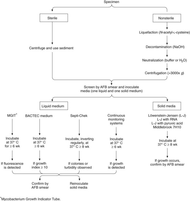
Processing to recover acid-fast bacilli from clinical specimens involves several complex steps, each of which must be carried out with precision. Specimens from sterile sites can be inoculated directly to media (small volume) or concentrated to reduce volume. Other specimens require decontamination and concentration. A processing scheme is shown in Figure 43-1, and the procedures are explored in detail in the following discussions.


Stay updated, free articles. Join our Telegram channel

Aug 25, 2016 | Posted by in MICROBIOLOGY | Comments Off on Mycobacteria

Full access? Get Clinical Tree

Get Clinical Tree app for offline access