Lined by mucinous columnar epithelium with focal cuboidal, flattened, or papillary areas
May have gastric or intestinal metaplasia
Varying degrees of dysplasia may be present
Densely cellular ovarian-like stroma positive for ER, PR, and inhibin
• MCN with associated invasive carcinoma
Most arise from preexisting MCN
Invasion of underlying stroma by malignant glands or single cells
Top Differential Diagnoses
• Cystic variant of biliary intraductal papillary neoplasm
• Solitary bile duct cysts
• Ciliated hepatic foregut cyst
• Endometrial cyst
Diagnostic Checklist
• Multilocular cystic neoplasm lined by mucinous epithelial cells with underlying ovarian-type stroma
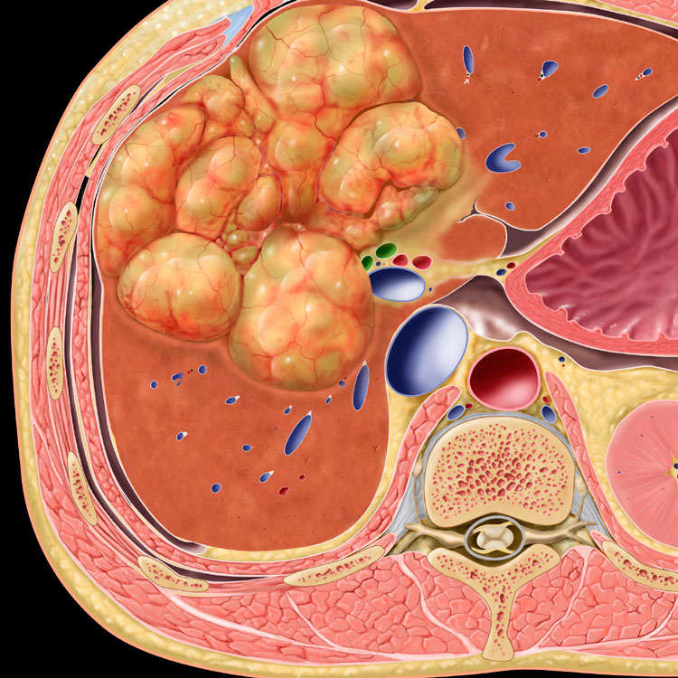
Schematic Representation Graphic shows a lobulated, complex cystic mass with a vascularized wall, areas of solid growth, and well-defined septa typical of mucinous cystic neoplasm (MCN) of the liver.
TERMINOLOGY
Abbreviations
• Mucinous cystic neoplasm (MCN)
Definitions
• Cystic biliary neoplasm arising within liver or in extrahepatic biliary tree, including gallbladder
• Formerly known as biliary cystadenoma and cystadenocarcinoma
ETIOLOGY/PATHOGENESIS
Unknown
• May originate from müllerian remnants misplaced during embryogenesis
• Proliferation of endodermally derived epithelium and primitive mesenchyme stimulated by female sex hormone
CLINICAL ISSUES
Epidemiology
• Incidence
Rare; < 5% of cystic lesions of liver
• Age
Average: 40-50 years
• Sex
Almost exclusively occurs in women
Presentation
• Pain, mass, and occasionally jaundice
Some patients are asymptomatic
Laboratory Tests
• CA19-9 and CEA in cyst fluid helps differentiate between simple cyst and MCN
Treatment
• Surgical approaches
Complete resection
Prognosis
• Complete surgical resection should be curative
Incompletely resected tumor may recur or undergo malignant transformation
IMAGING
Ultrasonographic Findings
• Large, well-defined, multiloculated, anechoic mass with highly echogenic septations
• Mural or septal calcifications or fluid levels
CT Findings
• Nonenhanced CT
Large, well-defined, homogeneous, hypodense heterogeneous mass (cystic and hemorrhagic areas)
Only gold members can continue reading. Log In or Register to continue