Other metastatic small cell neuroendocrine tumors [should be CK20(-)]
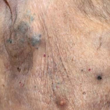
Clinical Photograph of Metastatic Melanoma Melanoma metastases are often blue-black in color. Typically, they are located in somewhat close proximity to the original primary. (Courtesy Yale Dermatology Residents’ Slide Collection.)
Metastatic Melanoma to Dermis This metastatic melanoma shows dermal invasion, with lack of epidermal involvement (note the presence of a thin grenz zone ). The tumor is composed of infiltrative nests and cords of atypical cells with melanin pigment in the dermis.
Metastatic Merkel Cell Carcinoma Other primary skin tumors can also metastasize to the skin. This is an example of metastatic Merkel cell carcinoma. There are islands composed of small, dark blue cells in the deep dermis.
Metastatic Sebaceous Carcinoma This is an example of a metastatic poorly differentiated sebaceous carcinoma to the skin of the cheek. Note the broad grenz zone separating the tumor from the epidermis .
TERMINOLOGY
Definitions
• Metastatic tumor originating from skin
CLINICAL ISSUES
Presentation
• Melanoma
Cutaneous metastases
– Nodule or papule, often pigmented, may be ulcerated, multiple or solitary
– Rarely presenting sign of disease; occasionally primary tumor site cannot be determined
Nodal metastases
– Most commonly in regional, draining lymph nodes
Distant metastases
– Any site, including bone, gastrointestinal tract, lung, brain
Blue nevus-like melanoma
– Blue macule/papule, often near original site of primary melanoma
• Merkel cell carcinoma
Spreads to lymph nodes in up to 50-75% of cases
Distant metastasis (e.g., liver, lungs, bone, brain) in up to 30-50% of cases
• Squamous cell carcinoma (SCC)
Nonspecific nodules, often pink to red, rarely zosteriform pattern
• Basal cell carcinoma (BCC)
Very rarely metastasizes to other sites (lymph nodes, bone, parotid, lungs, other internal organs)
Usually only in very large, deeply invasive, recurrent tumors
• Dermatofibrosarcoma protuberans (DFSP)
Essentially only fibrosarcomatous cases; often history of multiple local recurrences
Rarely metastasizes
– Lung most common site; also lymph nodes, bone, soft tissue
• Many other cutaneous malignancies may metastasize
Adnexal carcinomas
– Sebaceous carcinoma: Relatively high incidence of metastasis
Only gold members can continue reading. Log In or Register to continue


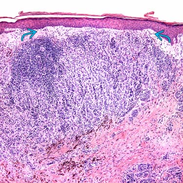
 ). The tumor is composed of infiltrative nests and cords of atypical cells with melanin pigment in the dermis.
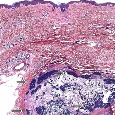
 composed of small, dark blue cells in the deep dermis.
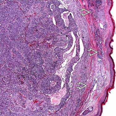
 .
.


