The cut surface shows numerous variably sized cystic spaces  admixed with solid areas. Minimal uninvolved liver tissue is present at the resection margin
  .
.
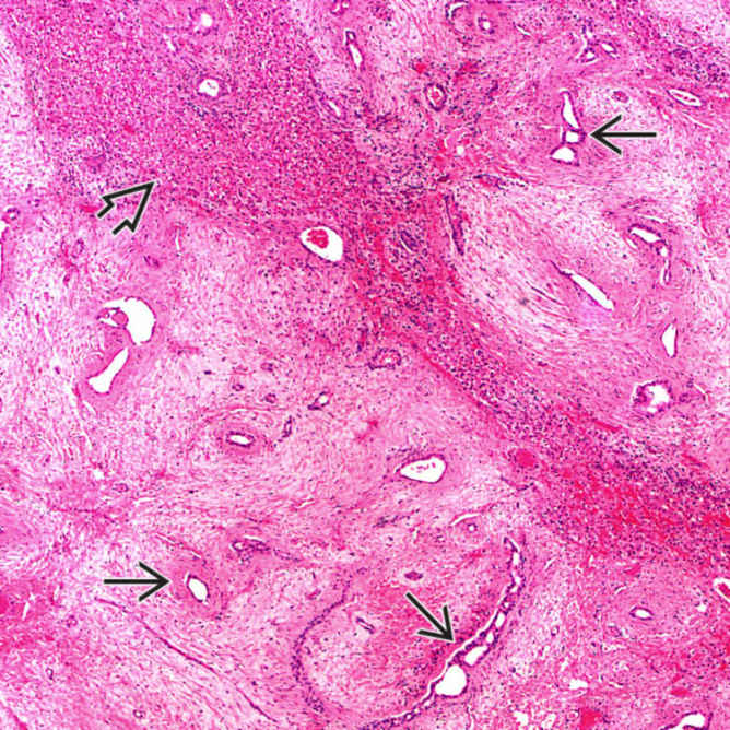
Low-power view shows numerous bile ducts
 in a loose, myxoid mesenchymal stroma. Note the presence of a large island of normal-appearing hepatocytes
  in the tumor.
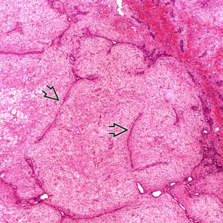
Compressed and branching bile ducts within collagenous stroma have a ductal plate malformation-like pattern
 .
.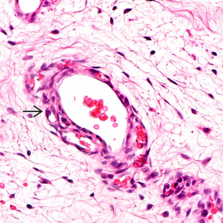
High-power view shows scattered stellate mesenchymal cells loosely embedded in an edematous stroma. Note the presence of blood vessels
 .
.


CLINICAL ISSUES
Epidemiology


Stay updated, free articles. Join our Telegram channel

Full access? Get Clinical Tree


