Meningitis, Encephalitis, and Other Infections of the Central Nervous System


Meningitis, Encephalitis, and Other Infections of the Central Nervous System


Objectives


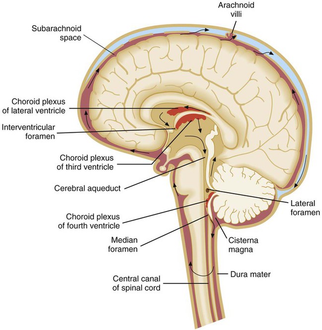
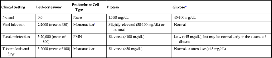
1. Describe the anatomy of the central nervous system (CNS), and list the anatomic structures that compose it.


2. Define meninges; name the three separate layers and describe their function.


3. Define the cerebrospinal fluid, and list the functions of the cerebrospinal fluid (CSF).


4. Describe the routes of infection for the central nervous system.


5. Explain the host defense mechanisms that protect the central nervous system from infection.


6. Define meningitis, and describe the two major types of meningitis including the etiologic agents.


7. When discussing the etiology of acute meningitis, explain the host predisposing factors for the neonate and identify the most commonly associated bacterial pathogens.


8. Discuss how the advent of the Hib vaccine in the United States has helped to prevent pediatric cases of meningitis.


9. Compare acute and chronic meningitis; outline the distinguishing symptoms and CSF findings for each, including cell counts and chemistry laboratory results.


10. Explain the disease processes for encephalitis and meningoencephalitis.


11. Discuss two ways that meningoencephalitis infections, brain abscesses, or other CNS infections are caused by parasites; identify the associated infecting organisms and the population of patients at increased risk for developing these conditions.


12. Explain the collection, transport, and specimen storage requirements for CSF; include specimen processing and the appropriate distribution of specimen throughout the laboratory.


13. List the culture media used to identify the causative agent of meningitis in bacterial, mycobacterial, and fungal infections; what incubation conditions are required for each type of organism?


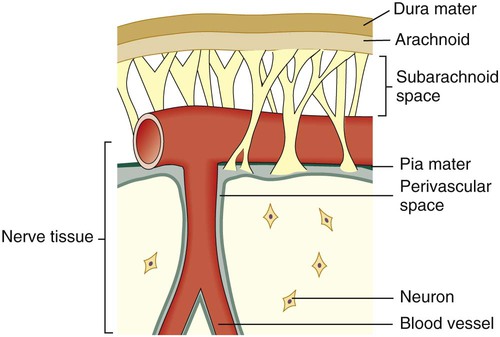
General Considerations


Anatomy


Diagnosis of an infection involving the central nervous system (CNS) is of critical importance. Most clinicians consider infection in the CNS to be a medical emergency. An understanding of the basic anatomy and physiology of the CNS is helpful for the microbiologist to ensure appropriate specimen processing and interpretation of laboratory results.


Coverings and Spaces of the CNS


The central nervous system consists of the brain and the spinal cord. Because of the vital and essential role of the CNS in the body’s regulatory processes, the brain and spinal cord have two protective coverings: an outer covering consisting of bone and an inner covering of membranes called the meninges. The outer bone covering encases the brain (i.e., cranial bones or skull) and spinal cord (i.e., the vertebrae). The meninges is a collective term for the three distinct membrane layers surrounding the brain and spinal column:


The pia mater and the arachnoid membrane are collectively called the leptomeninges. The portion of the arachnoid that covers the top of the brain contains arachnoid villi, which are special structures that absorb the spinal fluid and allow it to pass into the blood.


Between and around the meninges are spaces that include the epidural, subdural, and subarachnoid spaces. The relative location of the meninges and spaces to one another in the brain are depicted in Figure 71-1. The location and nature of the meninges and spaces are summarized in Table 71-1.


Cerebrospinal Fluid


Cerebrospinal fluid (CSF) surrounds the brain and spinal cord and has several functions. The CSF provides cushioning and buoyancy for the bulk of the brain, reducing the effective weight of the brain by a factor of 30. CSF carries essential metabolites into the neural tissue and cleanses the tissues of wastes as it circulates around the brain, ventricles, and spinal cord. Every 3 to 4 hours, the entire volume of CSF is exchanged. In addition to these functions, CSF provides a means by which the brain monitors changes in the internal environment.


CSF is found in the subarachnoid space (see Table 71-1) and within cavities and canals of the brain and spinal cord. There are four large, fluid-filled spaces within the brain referred to as ventricles. Specialized secretory cells, called the choroid plexus, produce CSF. The choroid plexus is located centrally within the brain in the third and fourth ventricles. Approximately 23 mL of CSF are contained within these ventricles in an adult. The fluid travels around the outside areas of the brain within the subarachnoid space, driven primarily by the pressure produced initially at the choroid plexus (Figure 71-2). By virtue of its circulation, chemical and cellular changes in the CSF may provide valuable information about infections within the subarachnoid space.


Routes of Infection


One of the most important defense mechanisms of the CNS is the blood-brain barrier. The blood-brain barrier functions to maintain homeostasis in the brain through restricting the flow of chemical constituents from the blood to the CNS. In order for the CNS to become infected with bacteria, parasite, or virus, the blood-brain barrier must be penetrated.


Organisms may gain access to the CNS through several primary routes:


• Hematogenous spread: followed by entry into the subarachnoid space through the choroid plexus or through other blood vessels of the brain. This is the most common route of infection for the CNS.


• Direct spread from an infected site: the extension of an infection close to or contiguous with the CNS can occasionally occur; examples of such infections include otitis media (infection of middle ear), sinusitis, and mastoiditis.


• Anatomic defects in CNS structures: anatomic defects as a result of surgery, trauma, or congenital abnormalities can allow microorganisms easy and ready access to the CNS.


• Travel along nerves leading to the brain (direct intraneural): the least common route of CNS infection caused by organisms such as rabies virus, which travels along peripheral sensory nerves, and herpes simplex virus.


Diseases of the Central Nervous System


Meningitis


Infection within the subarachnoid space or throughout the leptomeninges is called meningitis. Based on the host’s response to the invading microorganism, meningitis is divided into two major categories: purulent and aseptic meningitis.


Purulent Meningitis.

A patient with purulent meningitis typically has a marked, acute inflammatory exudative cerebral spinal fluid containing large numbers of polymorphonuclear cells (PMNs). Frequently, the underlying CNS tissue, in particular the ventricles, may be involved. If the ventricles become involved, this process is referred to as ventriculitis. Bacterial organisms are usually the cause of these infections.


Pathogenesis.

The outcome of a host-microbe interaction depends on the characteristics of both the host and the microorganism. As previously indicated, an important host defense mechanism within the CNS is the blood-brain barrier; this barrier involves the choroid plexus, arachnoid membrane, and the cerebral microvascular endothelium. The unique structural properties of the vascular endothelium, such as the continuous intercellular tight junctions, provide a barrier minimizing the passage of infectious agents into the CSF. The normal function of the vascular endothelium includes regulating the transport of nutrients in and out of the CSF, including low-molecular-weight plasma proteins, glucose, and electrolytes.


The host’s age and other underlying factors contribute to whether an individual is predisposed to the development of infectious meningitis. Neonates have the highest infection rate for meningitis, because of the immature neonatal immune system, the increased permeability of the blood-brain barrier in newborns, and the presence of colonizing bacteria in the female vaginal tract that can pass to the infant during childbirth. The most common bacterial pathogens responsible for meningitis in newborns are group B streptococci, Escherichia coli, and Listeria monocytogenes. Prior to the advent of the Hib vaccine in the United States in 1985, Haemophilus influenza type b (Hib) was a common cause of meningitis in children 4 months to 5 years of age. Because of the incorporation of Hib into childhood immunization programs, childhood Hib disease has dramatically declined.


Among young adults, Neisseria meningitidis is typically the agent that is associated with meningitis. N. meningitidis has been identified in epidemics among young adults in crowded conditions (e.g., military recruits and college dormitory mates). There are two meningococcal vaccines (vaccines for N. meningitidis) available in the United States. The meningococcal polysaccharide vaccine (MPSV4) is used for individuals older than 55 years of age, and the meningococcal conjugate vaccine (MCV4) is used for adolescents. Streptococcus pneumoniae is frequently the cause of meningitis in young children and the elderly; often this meningitis develops from bacteremia or from infection of the sinuses or middle ear. There are two pneumococcal vaccines (vaccines for S. pneumoniae) that are recommended currently in the United States. The pneumococcal conjugate vaccine (PCV13) protects against infection from 13 different serotypes of S. pneumoniae and is used for vaccination of children and adults. The second vaccine, pneumococcal polysaccharide vaccine (PPSV), provides protection from 23 serotypes of S. pneumoniae, including those associated with serious life-threatening infections. This vaccine is recommended for adults 65 years of age and older or anyone over the age of 2 who has long-term health problems or is immunocompromised.


Because the respiratory tract is the primary portal of entry for many etiologic agents of meningitis, factors that predispose adults to meningitis are often the same factors that increase the likelihood for the development of pneumonia or other respiratory tract colonization or infection. Alcoholism, splenectomy, diabetes mellitus, prosthetic devices, and immunosuppression contribute to increased risk. Finally, patients with prosthetic devices, particularly CNS and ventriculoperitoneal shunts, are at increased risk for developing meningitis.


For organisms to reach the CNS (primarily by the blood-borne route), host defense mechanisms must be overcome. Most cases of meningitis are a result of bacteria that share a similar pathogenesis. The successful meningeal pathogen must first sequentially colonize and cross host mucosal epithelium, then enter and thrive within the bloodstream. The most common causes of meningitis possess the ability to evade host defenses at each of these levels. For example, clinical isolates of Streptococcus pneumoniae and N. meningitidis secrete IgA proteases capable of destroying the host’s secretory IgA, thereby facilitating bacterial attachment to the epithelium. In addition, all of the most common etiologic agents of bacterial meningitis possess an antiphagocytic capsule that allows the organisms to evade destruction by the host immune system.


Organisms appear to enter the CNS by interacting and subsequently breaking down the blood-brain barrier at the level of microvascular endothelium. One of the least understood processes in the pathogenesis of meningitis is how organisms cross this barrier into the subarachnoid space. Nevertheless, there appear to be specific bacterial surface components, such as pili, polysaccharide capsules, and lipoteichoic acids, that facilitate adhesion of the organisms to the microvascular endothelial cells and subsequent penetration into the CSF. Organisms can enter (1) through loss of capillary integrity by disrupting tight junctions of the blood-brain barrier, (2) through transport within circulating phagocytic cells, or (3) by crossing the endothelial cell lining within endothelial cell vacuoles. After gaining access, the organism multiplies within the CSF, a site initially free of antimicrobial antibodies or phagocytic cells.


Acute.

Symptoms of acute meningitis include fever, stiff neck, headache, nausea and vomiting, neurologic abnormalities, and change in mental status.


In acute bacterial meningitis, the CSF usually contains large numbers of inflammatory cells (>1000/mm3), primarily polymorphonuclear cells (PMNs). The CSF shows a decreased glucose level relative to the serum glucose level and an increase in protein concentration. In a healthy individual, the normal CSF glucose level is 0.6 of the serum glucose level and ranges from 45 to 100 mg/dL; the CSF protein range in an adult is 15 to 50 mg/dL; newborn CSF protein ranges run as high as 170 mg/dL with an average of 90 mg/dL.


The sequelae of acute bacterial meningitis in children are frequent and serious. Seizures can occur in 20% to 30% of patients, and other neurologic changes are common. Acute sequelae include cerebral edema, hydrocephalus, cerebral herniation, and focal neurologic changes. Permanent deafness can occur in 10% of children who recover from bacterial meningitis. Other subtle physiologic and psychological sequelae may also follow an episode of acute bacterial meningitis.


Chronic.


Chronic meningitis can often occur in patients who are immunocompromised, although this is not always the case. Patients experience an insidious onset of disease, with some or all of the following symptoms: fever, headache, stiff neck, nausea and vomiting, lethargy, confusion, and mental deterioration. Symptoms may persist for a month or longer before treatment is sought. The CSF usually manifests an abnormal number of white blood cells (usually lymphocytic), elevated protein, and decrease in glucose content (Table 71-2). The pathogenesis of chronic meningitis is similar to that of acute disease.


Epidemiology/Etiologic Agents-Acute Meningitis.

The etiology of acute meningitis depends on the age of the patient. Most cases in the United States occur in children younger than 5 years of age. Before 1985, H. influenzae type b H. influenzae type b was the most common infectious agent in children between 1 month and 6 years of age within the United States. Ninety-five percent of all cases were due to H. influenzae type b, Neisseria meningitidis, and Streptococcus pneumoniae. In 1985, the first Hib vaccine, a polysaccharide vaccine, was licensed for use in children 18 months of age or older but was not efficacious in children younger than 18 months. However, the widespread use of conjugate vaccine, Hib polysaccharide-protein conjugate, in children as young as 2 months of age has significantly affected the incidence of invasive H. influenzae type b disease; the total number of annual cases of H. influenzae disease in the United States have been reduced by 55% and the number of cases of H. influenzae meningitis by 94%. However, the risks for meningococcal and pneumococcal diseases resulting from agents other than H. influenzae have remained level. Children older than 6 years of age are less likely to develop meningitis, but the risk for meningitis infection increases when the child reaches early adulthood. As previously mentioned, neonates have the highest incidence of acute meningitis, with a concomitant increased mortality rate (as high as 20%). Organisms causing disease in the newborn are different from those that affect other age groups; many of them are acquired by the newborn during passage through the mother’s vaginal vault. Neonates are likely to be infected with, in order of incidence, group B streptococci, Escherichia coli, other gram-negative bacilli, and Listeria monocytogenes; occasionally other organisms may be involved. For example, Elizabethkingia meningoseptica has been associated with nursery outbreaks of meningitis. This organism is a normal inhabitant of water in the environment and is presumably acquired as a nosocomial infection.


Important causes of meningitis in the adult, in addition to the meningococcus in young adults, include pneumococci, Listeria monocytogenes, and, less commonly, Staphylococcus aureus and various gram-negative bacilli. Meningitis caused by the latter organisms results from hematogenous seeding from various sources, including urinary tract infections. The percentage of adults with nosocomial bacterial meningitis at large urban hospitals has been increasing. The various etiologic agents of chronic meningitis are listed in Box 71-1.


Aseptic Meningitis.

Aseptic meningitis is usually viral and characterized by an increase of lymphocytes and other mononuclear cells (pleocytosis) in the CSF; bacterial and fungal cultures are negative. (This is in contrast to bacterial meningitis, which is characterized by purulence and the polymorphonuclear [PMN] cell response in the CSF.) Aseptic meningitis is usually self-limiting with symptoms that may include fever, headache, stiff neck, nausea, and vomiting.


In addition to the increase of lymphocytes and other mononuclear cells in the CSF, the glucose level remains normal, whereas the protein CSF level may remain normal or be slightly elevated. Aseptic meningitis can also be a symptom for syphilis and some other spirochete diseases (e.g., leptospirosis and Lyme borreliosis). Stiff neck and CSF pleocytosis may also be associated with other disease processes, such as malignancy.


Encephalitis/Meningoencephalitis


Encephalitis is an acute inflammation of the brain parenchyma and is usually caused by direct viral invasion. Concomitant meningitis occurring with encephalitis is known as meningoencephalitis, and the cellular infiltrate present in the CSF is typically lymphocytic rather than polymorphonuclear cells.


The host response to these CNS infections can differ somewhat from those associated with purulent or aseptic meningitis. Early in the course of viral encephalitis, or when considerable tissue damage occurs as a part of encephalitis, the nature of the inflammatory cells found in the CSF may be no different from that associated with bacterial meningitis; cell counts, however, are typically much lower.


Viral.


Viral encephalitis, which cannot always be distinguished clinically from meningitis, is common in the warmer months. The primary agents are enteroviruses (coxsackie viruses A and B, echoviruses), mumps virus, herpes simplex virus, and arboviruses (West Nile virus, togavirus, bunyavirus, equine encephalitis, St. Louis encephalitis, and other encephalitis viruses). Other viruses—such as measles, cytomegalovirus, lymphocytic choriomeningitis, Epstein-Barr virus, hepatitis, varicella-zoster virus, rabies virus, myxoviruses, and paramyxoviruses—are less commonly encountered. Any preceding viral illness and exposure history are important considerations in establishing a cause by clinical means. Since 1999, with the first debut of West Nile in the United States, the West Nile virus has been an important consideration in the diagnosis of viral encephalitis. The Centers for Disease Control and Prevention (CDC) reports that the incidence of West Nile infection peaked in 2003 with 9862 cases of West Nile infection; 2860 were reported cases of meningitis and encephalitis, resulting in 264 deaths. Since then the rates of infection have dropped: human cases reported to the CDC in 2010 were significantly lower with 1021 total reported cases of West Nile; 629 were neuroinvasive cases resulting in 57 deaths; a state-by-state breakdown of the disease incidence is outlined in Table 71-3. In 2012, a deadly resurgence of West Nile virus occurred, including neuroinvasive and non-neuroinvasive, for a total of 4531 cases through mid-October, according to the CDC.


TABLE 71-3


Final 2010 West Nile Virus Human Infections in the United States*


State Neuroinvasive Disease Cases Non-neuroinvasive Disease Cases Total Cases Deaths Presumptive Viremic Donors*
Alabama 1 2 3 0 0
Arizona 107 60 167 15 31
Arkansas 6 1 7 1 0
California 72 39 111 6 24
Colorado 26 55 81 4 1
Connecticut 7 4 11 0 5
District of Columbia 3 3 6 0 0
Florida 9 3 12 2 1
Georgia 4 9 13 0 1
Idaho 0 1 1 0 0
Illinois 45 16 61 4 5
Indiana 6 7 13 1 0
Iowa 5 4 9 2 1
Kansas 4 15 19 0 1
Kentucky 2 1 3 1 7
Louisiana 20 7 27 0 7
Maryland 17 6 23 2 0
Massachusetts 6 1 7 0 1
Michigan 25 4 29 3 2
Minnesota 4 4 8 0 1
Mississippi 3 5 8 0 2
Missouri 3 0 3 0 0
Nebraska 10 29 39 2 10
Nevada 0 2 2 0 0
New Hampshire 1 0 1 0 0
New Jersey 15 15 30 2 0
New Mexico 21 4 25 1 6
New York 89 39 128 4 16
North Dakota 2 7 9 0 0
Ohio 4 1 5 0 0
Oklahoma 1 0 1 0 1
Pennsylvania 19 9 28 0 0
South Carolina 1 0 1 0 0
South Dakota 4 16 20 0 0
Tennessee 2 2 4 0 1
Texas 77 12 89 6 14
Utah 1 1 2 0 3
Virginia 4 1 5 1 2
Washington 1 1 2 0 0
Wisconsin 0 2 2 0 1
Wyoming 2 4 6 0 0
Totals 629 392 1021 57 144

Only gold members can continue reading. Log In or Register to continue

Stay updated, free articles. Join our Telegram channel

Aug 25, 2016 | Posted by in MICROBIOLOGY | Comments Off on Meningitis, Encephalitis, and Other Infections of the Central Nervous System

Full access? Get Clinical Tree

Get Clinical Tree app for offline access