Fig. 31.1
Keratotic area on the inner ankle of a patient with lupus erythematosus with a 20-year history of immune suppression with numerous medications. The biopsy showed a squamous cell carcinoma. The important feature is the benign appearance, indicating the importance of being highly suspicious of tumors in this group of immune-suppressed patients
NMSCs that are increased include squamous cell carcinoma (10-to-1 basal-to-squamous cell cancer is reversed in long-term immunosuppressed patients), keratoacanthoma (Fig. 31.2), basal carcinoma, and merkel cell carcinoma. Precancerous tumors that are increased are porokeratosis (Fig. 31.3), actinic keratosis (Fig. 31.4), Bowen’s disease, and warts in both genital and non-genital (Fig. 31.5) locations.
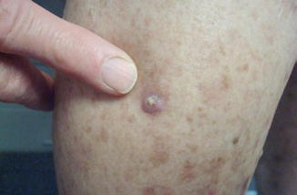
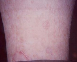
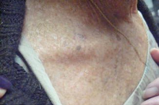
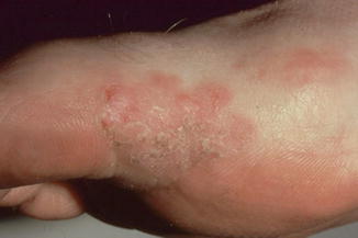
Fig. 31.2
Keratoacanthoma on the inner calf, with a rapidly growing pink tumor. It is a symmetric, circular, pink tumor with a central keratotic core. The patient had been on azathioprine for decades
Fig. 31.3
Porokeratosis on the pretibial area in center of figure (smaller lesion) and located in the lower left of the photo (larger lesion that is cut off). The thin linear keratotic rim around the tumor is the diagnostic sign. The commonest type of porokeratosis is disseminated actinic porokeratosis. In the immune-suppressed the porokeratosis tend to be asymmetric in distribution, thicker, less common, and more likely to become a squamous cell carcinoma
Fig. 31.4
Keratotic brown tumor on right supraclavicular area that is ill-defined, with a subtle pink base that was an actinic keratosis; in a patient who was immune-suppressed from scleroderma. There is a brown well-demarcated stuck-on tumor below the actinic keratosis and to the right of the actinic keratosis, near the patient’s necklace. These two tumors are seborrheic keratoses
Fig. 31.5
An eczematous plaque on the inner edge of the base of the great toe. Bowen’s disease often mimics a dermatitis such as eczema or psoriasis
PUVA
A landmark paper published in 1974 supported the use of psoralen plus ultraviolet A (PUVA) as an effective treatment for psoriasis, as well as more recently for cutaneous T-cell lymphoma stage IA and IB, eczema, vitiligo, graft-versus-host disease, and atopic dermatitis. Psoralen is a naturally occurring phototoxic compound that absorbs light photons and alters DNA and cell components. It may be applied topically or taken orally 1–2 h before UVA application. Psoralen initially penetrates into the cell between DNA, and upon activation via UVA radiation, DNA base pairs are cross-linked, leading to cell apoptosis, mutagenesis, and photocarcinogenesis. More than just inhibiting cell proliferation, PUVA has immunomodulatory properties, such as changing cytokine expressions and functionality of antigen presenting cells.
Since PUVA is mutagenic and exhibits immunosuppressive properties in the skin, it has always been considered a human carcinogen. From 1975 to 2005, a major clinical trial of 1380 psoriatic patients on PUVA was initiated to study the long-term safety profile of PUVA. Results showed that there is a dose-dependent increase in the risk of SCC and moderate increase in risk of BCC at increasing PUVA radiation, which persisted even after cessation of treatments. Patients who had 350+ PUVA treatments had a six-fold increased risk of developing SCC compared with patients who had fewer than 50 treatments. High UVB was associated with an increased risk of BCC. The locations of PUVA-induced SCC also differs from UV-induced SCC, with over half the lesions on the lower extremities versus the more common sun-exposed regions on the head and neck. Men exposed to PUVA treatments are also 53 times more likely to develop invasive scrotal or penile squamous cell carcinomas than the general Caucasian population. A Swedish retrospective study with a 16-year follow-up found that patients who used PUVA have increased risks for developing cutaneous SCC with a relative risk of 5.6 for men and 3.6 for women.
Prior to the 1990s, many retrospective reviews did not appreciate the increased risk of skin cancer with PUVA therapy. This may be due to short follow-up periods (skin cancer changes take years) or because many psoriatic patients had previously received additional carcinogenic treatments including coal tar, radiotherapy, and arsenic. Interestingly, it has been observed that the increased risk of skin cancer after PUVA treatments is higher in U.S. patients than in European patients. This may be related to skin phototype differences (I–II in US vs. III–IV in Europe) or differences with the treatment approach in which Europe is more aggressive with higher single-dose treatments versus the U.S., which is more conservative, with a lower dose but longer exposure time. PUVA is an effective psoriatic treatment, but its effectiveness must be weighed against the increased risk of developing SCC.
Immunomodulatory Drugs—Thiopurines
Azathioprine (AZA) and 6-mercaptopurine (6-MP) are steroid-sparing agents that are commonly used for inducing and maintaining remission in Crohn’s disease, ulcerative colitis, and autoimmune diseases such as lupus erythematosus. These have been used in solid-organ transplant patients in the past, but now less commonly used. AZA is a prodrug that is converted nonenzymatically to 6-MP in the body. 6-MP is then metabolized by the liver and gut via one of three enzymes: thiopurine-S-methyltransferase (TPMT), xanthine oxidase, and hypoxanthine-guanine-phosphoribosyltransferase. The active metabolite, 6-thioguanine (6-TG) nucleotides, inhibits purine synthesis and eventually downregulates DNA and RNA synthesis. It also inhibits T- and B- lymphocytes proliferation, decreasing their production and causing apoptosis of T-cells.
Many retrospective studies showed causal relationships between uses of thiopurine and increased risk of NMSCs. It is thought that 6-TG inserts into the skin’s DNA and lowers the minimal erythema dose for UVA, but not UVB, light. DNA with 6-TG is extremely photosensitive (Fig. 31.6), in contrast to normal DNA bases that do not absorb UVA light to a significant degree, which promotes DNA misregulations including DNA breakage, crosslinking, and oxidation of nucleotides. This advances to formation of reactive oxygen species (ROS) and increased risk of developing skin cancer.
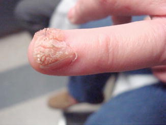
Fig. 31.6
Severe verrucae with destruction of the nail plate on the digit of a leukemia patient undergoing chemotherapy. Subungual warts can become squamous cell carcinomas in the immune-suppressed patient and a biopsy, as was done in this patient, is necessary to rule out transformation to a squamous cell carcinoma
A recent study showed an association between thiopurine usage in IBD patients and NMSC carry a relative risk of 4.9. Patients who may be more genetically susceptible, including those with a TPMT deficiency, should monitor their skin changes more frequently. A decreased level of TPMT may reduce metabolic clearance of thiopurines, leading to prolonged exposures inside the body and increased risks for NMSC. The patient’s past and family history are important, and it may be worthwhile to test for this gene’s commonly inherited polymorphisms, considering that as many as 10 % of the population carries a low-activity variant allele.
AZA is also widely used as an immunosuppressant in solid-organ transplant patients. Previous studies demonstrated post-transplant patients on multiple immunosuppressants have a 200-fold increased risk of developing NMSC. Data have suggested that transplant patients who continuously develop NMSC may switch from AZA to a possible lower-risk drug, such as mycophenolate mofetil or sirolimus.
The risk of developing NMSC with thiopurine usage is highest among Caucasian patients. There is an addictive effect, with previous UV light exposure combined with thiopurine and increased NMSC risk. Good sun-protective techniques should begin in childhood, including using sunscreen SPF 45+ and wearing protective headgear with a large brim.
DMARDs—Methotrexate, Biologic agents
Methotrexate (MTX) is a commonly prescribed disease-modifying antirheumatic drug (DMARD) for treating rheumatoid arthritis (RA), psoriasis, psoriatic arthritis, atrophic dermatitis, and other inflammatory conditions. MTX inhibits folic acid synthesis and alters the building blocks for DNA and RNA production. Early studies showed that psoriatic patients over 65 years old have a three-fold increased risk for developing lymphomas. A follow-up large retrospective study with more than 150,000 patients showed that psoriasis is associated with increased risks for Hodgkin’s lymphoma (HL) and cutaneous T-cell lymphoma (CTCL). Those that have more severe psoriatic conditions have the strongest relative risks for developing CTCL. The pathophysiology of abnormal T-cell proliferation and signaling in psoriasis may explain the increased risk to CTCL, but immunomodulating treatments may also provoke lymphoma development. Given the positive association between psoriasis and lymphoma, the absolute risk of lymphoma is still relatively low, such that it only affects a small subset of psoriatic patients.
Stay updated, free articles. Join our Telegram channel

Full access? Get Clinical Tree


