Massive Localized Lymphedema in Morbid Obesity
Steven D. Billings, MD
Key Facts
Clinical Issues
Seen in morbidly obese patients (> 300 lbs)
Macroscopic Features
Overlying skin has “peau d’orange” appearance
Pronounced fibrous tissue septa in subcutaneous fat
Cyst formation common
Cut section weeps serous fluid
Microscopic Pathology
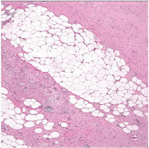
Expanded subcutaneous septa
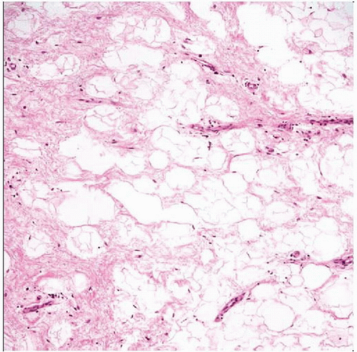
Prominent edema fluid
No pleomorphic hyperchromatic cells
Absence of plexiform vasculature, prominent myxoid stroma, and lipoblasts of myxoid liposarcoma
Preserved lobular architecture of subcutaneous fat
Reactive capillary vascular proliferation often present at interface of subcutaneous septa and lobules
 Hematoxylin & eosin shows cellular fibrous septa of variable width within the subcutaneous adipose tissue. No nuclear pleomorphism is seen in either the fibrous septa or fat. |
TERMINOLOGY
Definitions
Pseudoneoplastic process related to localized lymphatic obstruction
CLINICAL ISSUES
Presentation
Painless mass
Seen in morbidly obese patients (> 300 lbs)
Most common on medial thigh
Can occur in other locations such as abdomen and axilla
Prognosis
Benign
Recurrence or persistence common
IMAGE FINDINGS
CT Findings
Soft tissue streaking due to expanded subcutaneous septa
Cysts
No discrete mass
MACROSCOPIC FEATURES
Stay updated, free articles. Join our Telegram channel

Full access? Get Clinical Tree


