QUESTION 37.4
A. Biliary cystadenoma
B. Hepatoblastoma
C. Infantile hemangioma
D. Mesenchymal hamartoma
E. Polycystic liver disease
5. The autopsy of a 40-year-old woman reveals hemoperitoneum and a subcapsular hemorrhagic mass in the right hepatic lobe. The mass is partially encapsulated, and the adjacent liver parenchyma appears compressed. This picture shows its histologic composition. Conspicuous thick-walled vessels are scattered throughout the lesion, but there are no identifiable central veins or portal tracts. Which of the following is the most important predisposing factor for this lesion?
QUESTION 37.5
A. Aflatoxin exposure
B. Bartonella henselae infection
C. Hepatitis B virus infection
D. Oral contraceptive use
E. Thorotrast exposure
6. A hepatocellular adenoma (HCA) removed from the liver of a 45-year-old man shows prominent cytologic atypia. Which of the following immunohistochemical features is this HCA type most likely to display?
A. Lack of expression of liver fatty acid–binding protein (LFABP)
B. Nuclear expression of beta-catenin
C. Positivity for amyloid A–associated protein (SAA)
D. Positivity for LFABP, negative for beta-catenin and SAA
7. A well-demarcated subcapsular nodule is identified by MRI in the right hepatic lobe of a 35-year-old woman with abdominal discomfort. The lesion is removed. Its cut section is shown in this macroscopic photograph. Microscopic examination shows liver lobules with fatty changes separated by thick fibrous bands. The central scar contains thick-walled arteries. These gross and microscopic features are consistent with:
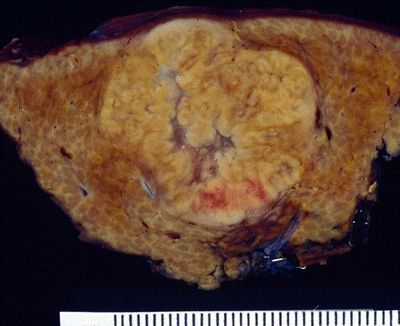
QUESTION 37.7
A. Early cirrhosis
B. Focal nodular hyperplasia
C. Hepatoblastoma
D. Liver adenoma
E. Nodular regenerative hyperplasia
8. A 50-year-old woman with rheumatoid arthritis presents with signs and symptoms of portal hypertension. An abdominal MRI is suggestive of liver cirrhosis. A needle biopsy reveals areas of congestion and atrophy alternating with areas of expanded lobules. Central veins are not seen. Which of the following mechanisms plays a crucial role in the pathogenesis of this disorder?
A. Drug-induced injury to hepatocytes
B. Drug-induced injury to sinusoids
C. Extrahepatic biliary injury
D. Hepatitis B virus infection
E. Microvascular alterations
9. Increasing utilization of screening and surveillance programs, combined with improved accuracy of imaging techniques, makes detection of small nodular lesions more common in cirrhotic livers. Some of these nodules may be cancer precursors. Which of the following nodular lesions has the highest likelihood to progress to hepatocellular carcinoma?
A. Dysplastic nodule, large cell change
B. Dysplastic nodule, small cell change
C. Regenerative nodule, monoacinar
D. Regenerative nodule, multiacinar (macroregenerative nodule)
10. Which of the following is the most reliable indicator of malignancy in a needle biopsy of a nodule within a cirrhotic liver?
A. Marked cellular atypia
B. Stromal invasion
C. Unpaired arteries
D. Vascular invasion
E. All of the above
11. The following is a list of factors or conditions that predispose to the development of hepatocellular carcinoma (HCC). In which of them does HCC occur constantly in noncirrhotic livers?
A. Alcohol abuse
B. Hepatitis B virus
C. Hepatitis C virus
D. Hereditary hemochromatosis
E. Hypercitrullinemia
A. Pseudoglandular
B. Scirrhous
C. Solid
D. Spindle cell
E. Trabecular
13. Which of the following immunohistochemical stains has a specificity for HCC that is close to 100%?
A. Arginase-1
B. Glypican-3
C. HepPar-1
D. MOC-31
E. Polyclonal CEA, canalicular pattern
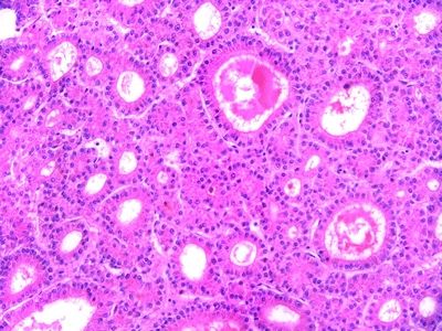
14. A patient undergoes partial hepatectomy for a neoplasm. The tumor consists of nodules of large cells with prominent nucleoli, separated by broad fibrohyaline bands, as shown in this picture. Which of the following is the most likely clinical setting associated with this tumor type?
QUESTION 37.14
A. Markedly elevated serum alpha-fetoprotein
B. A 20-year-old patient with chronic hepatitis B
C. A 30-year-old patient with normal liver
D. A 50-year-old patient with alcoholic cirrhosis
E. Elderly patient with hemochromatosis
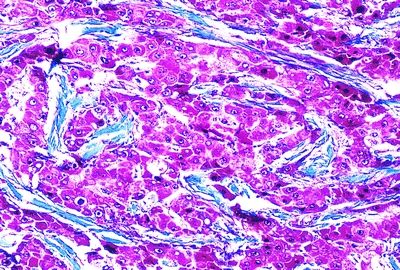
15. A 16-month-old baby is diagnosed with a liver tumor. On histologic examination, the tumor is characterized by a “light/dark” biphasic pattern, as shown in this low-power photomicrograph. This tumor is most likely a:
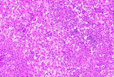
QUESTION 37.15
A. Bile duct hamartoma
B. Hemangioblastoma
C. Hepatoblastoma
D. Malignant mesenchymoma
E. Mesenchymal hamartoma
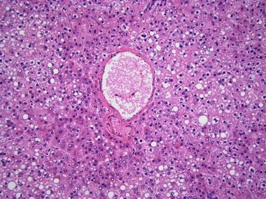
16. The cut surface of a liver removed at autopsy shows scattered white nodules in the liver parenchyma. Histologically, they consist of disorderly clusters of bile ductules lined by bland epithelial cells without identifiable connection with the bile system. These features are most consistent with:
A. Bile duct adenoma
Stay updated, free articles. Join our Telegram channel

Full access? Get Clinical Tree


