Glandular differentiation is often present; not as common as in benign mixed tumor of skin


• Infiltration of surrounding tissue, vascular invasion, and necrosis are helpful diagnostic features
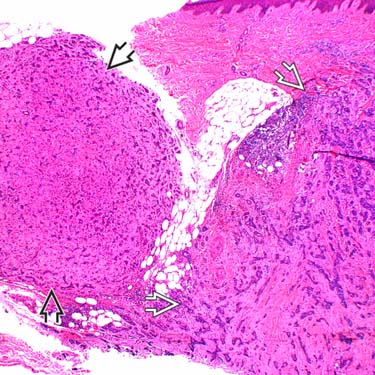
Malignant mixed tumor (MMT)
 arising in a longstanding benign mixed tumor (well-circumscribed nodule on the left
  ) shows a large, asymmetrical, lobular, and infiltrative tumor invading from the dermis into the subcutaneous tissue.
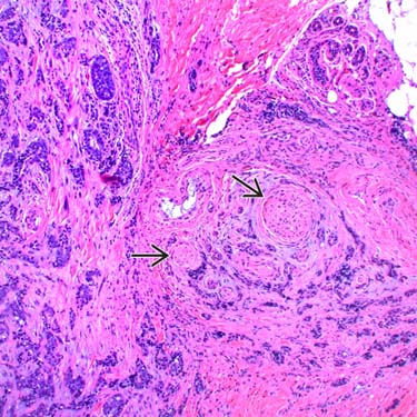
Marked infiltrative features with deep dermal and perineural
 invasion is observed.
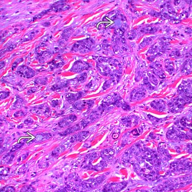
High magnification of an epithelial area shows infiltrative cords of cells, which demonstrate marked cytologic atypia with enlarged, hyperchromatic-staining nuclei surrounding small ductal lumina
 .
.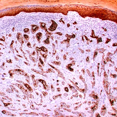
The epithelial component of the tumor is strongly highlighted by a HMWCK (CK5-CK6) stain.
CLINICAL ISSUES
Presentation

• Rarely, malignant transformation from mixed tumor of skin


Stay updated, free articles. Join our Telegram channel
Full access? Get Clinical Tree
