•
Lymphangioma circumscriptum (LC) (superficial cutaneous lymphangioma)
•
Lymphangiomatosis (LS) (generalized lymphangioma, systemic angiomatosis)
•
Cystic lymphangioma (cystic hygroma)
•
Deep lymphangioma (cavernous lymphangioma)
•
Proliferation of lymphatic vessels
Superficial (lymphangioma circumscription)
Deep (cavernous lymphangioma)
Diffusely involve most organ systems (LS)
•
Most cases are considered developmental or congenital malformations/hamartomas,
not true neoplasms
Maldevelopment during embryonic lymphangiogenesis most likely etiology
–
Leads to sequestered lymphatics that fail to communicate with normal lymphovascular system
May be due to maternal infections or substance abuse
LS considered congenital in most cases
•
Associated with genetic syndromes, including Turner syndrome (cystic hygroma), Noonan syndrome, Maffucci syndrome, trisomies 13, 18, 21
•
Mutations in
VEGFR-C ,
VEGFR3 ,
PROX1 ,
FOXC2 , and
SOX18 genes implicated
•
Rare acquired cases occur in adults
Likely associated with infection or trauma
•
Incidence
More common in children: Estimated 6% of benign childhood tumors
•
Age
Often present at birth or within first 2 years of life (~ 90% of cases)
LS usually presents within first 2 decades of life
•
Sex
Intraabdominal lymphangiomas have slight male predominance
LS has no gender predilection
•
Head and neck most common site for cystic lymphangiomas
Usually posterior triangle but can occur in anterior triangle
Also occur in axillae, abdomen, and internal organs
•
Cavernous type more frequent in oral cavity, upper trunk, limbs, and abdominal sites
Intraabdominal lymphangiomas occur in mesentery, omentum, and retroperitoneum
•
LC: Axillary folds, neck, and trunk are most common sites
•
LS: Can affect any organ system but often involves bones, soft tissues, and skin
•
Cystic mass lesion; may be superficial or deep
Typically presents as large, slow-growing, painless mass (deep lymphangioma) or as multiple small, grouped, superficial vesicular lesions (LC)
LS presents with numerous cystic lesions, both superficial and deep
•
Soft and fluctuant swellings on palpation
•
Intraabdominal cases may present with abdominal distension, mass on palpation
May also develop abdominal obstruction, volvulus, and infarction
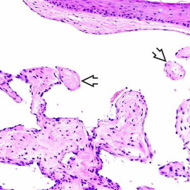
 with slightly fibrinous cores lined by small, hyperchromatic-staining endothelial cells.
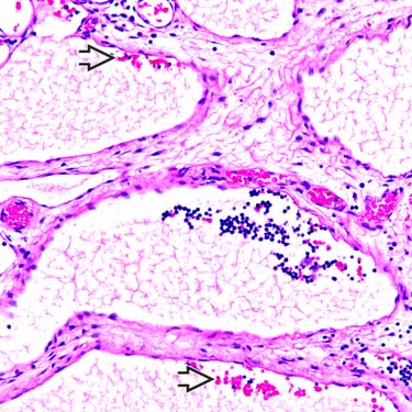
 .
.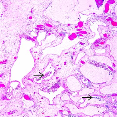
 within the lumina, along with fluid and erythrocytes.


 Typically presents as large, slow-growing, painless mass (deep lymphangioma) or as multiple small, grouped, superficial vesicular lesions (LC)


