QUESTION 17.3
A. Acquired immunodeficiency syndrome
B. Catscratch disease
C. Syphilis
D. Systemic lupus erythematosus
E. Toxoplasmosis
4. A young woman presents with fever, enlarged cervical lymph nodes, and leukopenia on the cell blood count. A lymph node biopsy reveals extensive areas of necrosis with nuclear debris surrounded by lymphocytes, histiocytes, and large numbers of plasmacytoid monocytes. Neutrophils are not present in the necrotic areas. Which of the following is the most likely diagnosis?
A. Angiofollicular hyperplasia (Castleman disease)
B. Catscratch disease
C. Histiocytic necrotizing lymphadenitis (Kikuchi-Fujimoto disease)
D. HIV infection
E. Syphilis
5. This photomicrograph shows the most characteristic set of histologic changes that develop in regional lymph nodes 2 to 3 weeks after a cat bite or scratch. Which of the following is it?
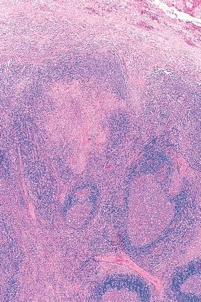
QUESTION 17.5
A. Capsulitis, subcapsular necrotizing granulomas, and follicular hyperplasia
B. Emperipolesis and expansion of sinuses by histiocytes
C. Follicular hyperplasia and interfollicular plasmacytosis
D. Germinal centers with ragged margins and marked interfollicular hyperplasia
E. Nonnecrotizing granulomas
6. The pattern of follicular hyperplasia seen in this lymph node is most characteristic of:
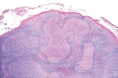
QUESTION 17.6
A. Acquired immunodeficiency syndrome
B. Catscratch disease
C. Syphilis
D. Systemic lupus erythematosus
E. Toxoplasmosis
7. A biopsy of a large mediastinal mass reveals the microscopic features detailed in these low-power and high-power photomicrographs (A and B), consisting of atretic lymphoid follicles, with a central hyalinized vessel surrounded by concentric layers of lymphocytes. Which of the following is true about this condition?
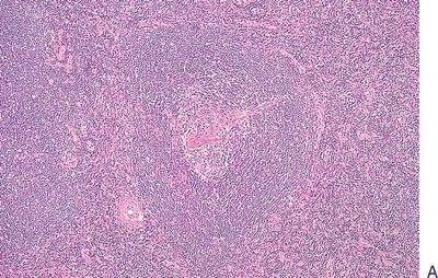
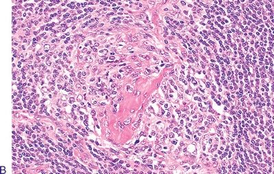
QUESTION 17.7
A. Human herpesvirus 8 (HHV-8) plays no pathogenetic role.
B. Necrosis is a constant feature of lymph node involvement.
C. Plasma cell population is monoclonal.
D. Plasma cell variant is more likely associated with systemic involvement.
E. Prognosis is the same for both localized and systemic forms.
8. A lymph node biopsy from a patient with fever, cervical lymphadenopathy, and splenomegaly reveals the histopathologic changes depicted in these photomicrographs, which consist of marked interfollicular expansion by a mixed cellular infiltrate with numerous immunoblasts, small lymphocytes, and few plasma cells. The follicular architecture is partially preserved. Which of the following is the most likely diagnosis?
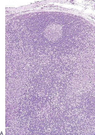
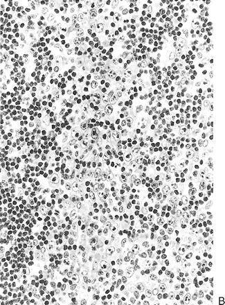
QUESTION 17.8
A. Angiofollicular hyperplasia
B. Angioimmunoblastic lymphoma
C. Catscratch disease
D. Immunoblastic lymphadenopathy
E. Infectious mononucleosis
9. A characteristic feature of sinus histiocytosis with massive lymphadenopathy (Rosai-Dorfman disease) is shown in this picture. Which feature is it?
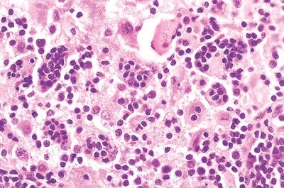
QUESTION 17.9
A. Emperipolesis
B. Interstitial Alcian blue–positive material
C. Interstitial proteinaceous deposits
D. Numerous immunoblasts
E. Melanin pigment
10. A young patient presents with localized cervical lymph node enlargement and fever. One of the lymph nodes is biopsied and reveals the changes shown in this picture. Which of the following features is frequently associated with the lymph node pathology in this condition?
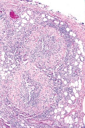
QUESTION 17.10
A. Amianthoid fibers
B. Obliterative vasculitis in perinodal vessels
C. Florid granulomatous inflammation
D. Hemophagocytosis
E. Slit-like vascular spaces
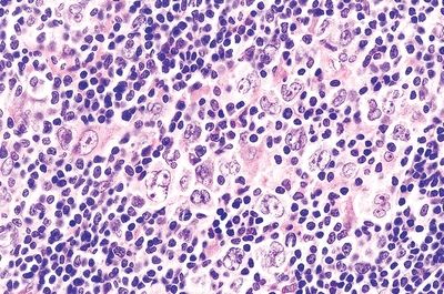
11. A 25-year-old man presents with cervical node enlargement. A biopsy of an affected lymph node shows effacement of lymph node architecture by a nodular growth, a thin rim of compressed normal node, and a predominantly small lymphocytic population with admixed histiocytes and scattered large cells like those seen in this picture. The latter are immunoreactive for CD45, CD20, CD79a, Bcl-6, and PAX-5 and negative for CD15. Which of the following is the Hodgkin lymphoma (HL) variant in this case?
QUESTION 17.11
A. Lymphocyte-depleted HL
B. Lymphocyte-rich classic HL
C. Nodular lymphocyte-predominant HL
D. Mixed cellularity HL
E. Nodular sclerosis HL
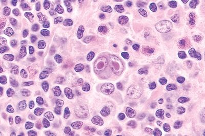
12. A biopsy of an enlarged cervical lymph node reveals a nodular growth pattern that effaces the nodal architecture, sparing an attenuated rim of residual lymphoid tissue. Within a cellular background rich in small lymphocytes, classic RS cells are identified. The latter express CD15, but not CD45. Which of the following is the HL variant in this case?
QUESTION 17.12
A. Lymphocyte-depleted HL
B. Lymphocyte-rich classic HL
C. Nodular lymphocyte-predominant HL
D. Mixed cellularity HL
E. Nodular sclerosis HL
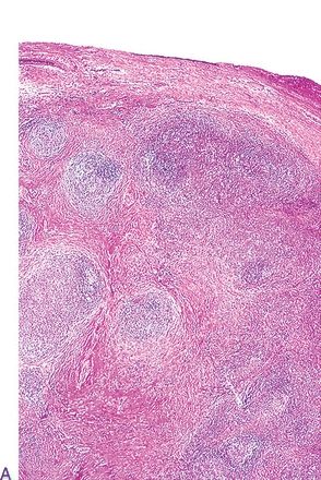
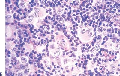
13. A lymph node biopsy in a case of suspected lymphoma reveals a nodular pattern of growth with broad bands of collagen, as shown in this low-power photomicrograph (A). A mercuric chloride–fixed section demonstrates a mixed lymphohistiocytic, plasmacellular, and eosinophilic infiltrate, along with the cells shown in this high-power photomicrograph (B). These cells are CD15 and CD30 positive and CD45 negative. Which of the following is the HL variant in this case?
QUESTION 17.13
A. Lymphocyte-depleted HL
B. Lymphocyte-rich classic HL
C. Nodular lymphocyte-predominant HL
D. Mixed cellularity HL
E. Nodular sclerosis HL
14. A lymph node biopsy reveals the cell population shown in this picture, including many classic RS cells. In this case, a diagnosis of mixed cellularity Hodgkin lymphoma (MCHL) is justified if:
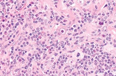
QUESTION 17.14
A. Broad bands of birefringent collagen result in a nodular pattern.
B. Classic Reed-Sternberg (RS) cells are rare.
C. Lacunar cells are abundant.
D. RS cells are easily found within an infiltrate of lymphocytes, histiocytes, plasma cells, and eosinophils.
E. RS cells are immunoreactive for CD45 but negative for CD15
15. Which of the following is true about lymphocyte-depleted Hodgkin lymphoma (LDHL)?
A. Accounts for more than 50% of HL cases.
B. Bone marrow involvement is rare.
C. Most patients present with localized cervical lymphadenopathy.
D. Reed-Sternberg (RS) cells are CD45+ and CD15–.
E. Some cases show diffuse fibrosis with PAS-positive material.
16. Which of the following immunophenotypes is characteristic of classical RS cells?
A. CD15+ CD30+ CD45−
B. CD15− CD30− CD45+
C. CD5+ CD10− CD23+
D. CD5− CD10+ CD43+
E. CD5− CD10+ Bcl-6+
17. A lymph node biopsy from a 60-year-old man with peripheral lymphocytosis and generalized lymphadenopathy reveals complete effacement of the nodal architecture, as shown in this picture. This is replaced by diffuse sheets of small lymphocytes with round nuclei and scanty cytoplasm. Mitotic figures are rare. Which of the following immunocytochemical, cytogenetic, or molecular findings would be consistent with this morphology?
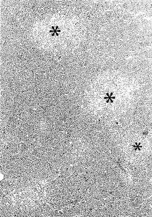
QUESTION 17.17
A. Immunoreactivity for CD5 and CD23
B. Immunoreactivity for CD10 and CD20, negativity for CD5
C. Strong immunoreactivity for surface immunoglobulin and CD43
D. Translocation t(8;14) with overexpression of c-myc
E. Translocation t(14;18) resulting in BCL-2 overexpression
18. A 65-year-old woman is found to have enlargement of multiple lymph nodes. Involvement of the spleen and liver is also documented. A lymph node biopsy reveals complete architectural effacement by a mixed population of lymphocytes, plasmacytoid lymphocytes, and plasma cells. Some of the plasmacytoid lymphocytes have nuclear inclusions highlighted by PAS staining, as shown in this picture. Which of the following features is associated with most cases of this lymphoproliferative disorder?
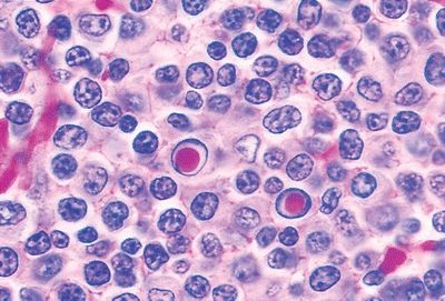
QUESTION 17.18
A. Bence Jones protein in the urine
B. Isolated bone mass
C. Primary amyloidosis
D. Richter syndrome
E. Waldenström macroglobulinemia
19. A 70-year-old man suffers from a lymphoma involving lymph nodes, bone marrow, spleen, and brain. Histologically, the lymph nodes display a vaguely nodular architecture, with striking expansion of the mantle zone around small residual follicles, as shown in this low-power photomicrograph (A). This high-power photomicrograph (B) demonstrates the cytologic features of the abnormal lymphocytic population. Which of the following genotypic abnormalities characterizes this type of lymphoma?
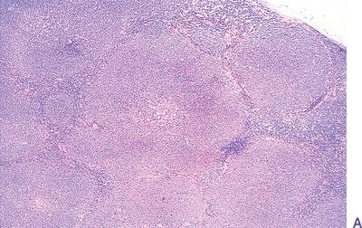
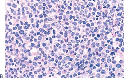
QUESTION 17.19
A. t(8;14)
B. t(11;14)
C. t(14;18)
D. Trisomy 12
E. Trisomy 18
20. This low-power photomicrograph shows a lymphoma composed of CD10-positive and CD5-negative lymphocytes with high levels of cytoplasmic IgG. Which of the following cytogenetic abnormalities is most likely to be present?
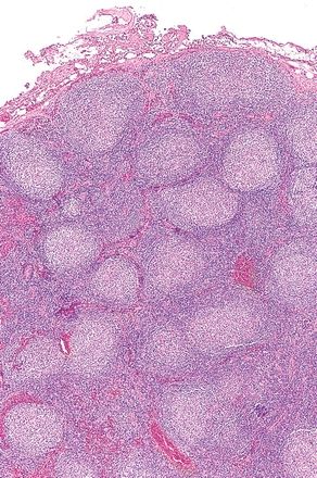
QUESTION 17.20
A. t(8;14)
B. t(11;14)
C. t(14;18)
D. Trisomy 12
E. Trisomy 18
21. Which of the following methods is used for grading follicular lymphomas?
A. Assessing degree of dysplasia of centrocytes
B. Counting centroblasts in 10 high-power fields
C. Counting centrocytes in 10 high-power fields
D. Estimating extent of follicular growth pattern
E. Estimating proportion of large transformed cells
22. This picture shows a lymphoma composed of neoplastic cells resembling monocytoid B cells. Which one is it?
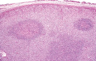
QUESTION 17.22
A. Follicular lymphoma
B. Hairy cell leukemia
C. Mantle cell lymphoma
D. Mastocytosis
E. Nodal marginal zone lymphoma
23. A 60-year-old man presents with rapidly enlarging axillary lymph nodes. A node biopsy shows the atypical lymphocytic population depicted in this photomicrograph. Neoplastic cells express CD19, CD20, CD79a, PAX-5, and CD10, while they are negative for CD5 and surface immunoglobulins. Which of the following is this type of lymphoma?
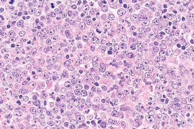
QUESTION 17.23
A. Burkitt lymphoma
B. Diffuse large B-cell lymphoma
C. Follicular lymphoma, high grade
D. Lymphoplasmacytic lymphoma
E. T-cell lymphoblastic lymphoma
For questions 24 through 32, match each description with the corresponding subtype of diffuse large B-cell lymphoma (DLBCL) listed below:
A. ALK positive
B. Arising in HHV-8-associated multicentric Castleman disease
Stay updated, free articles. Join our Telegram channel

Full access? Get Clinical Tree


