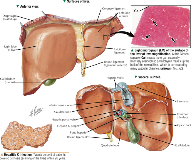
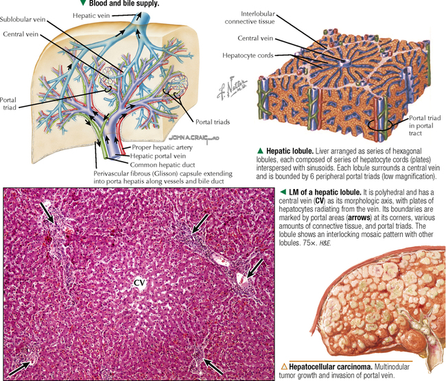
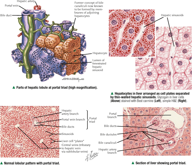
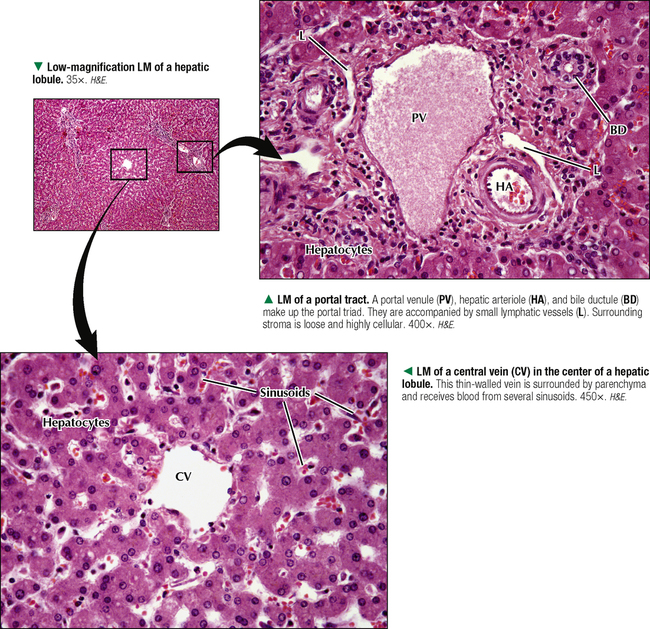
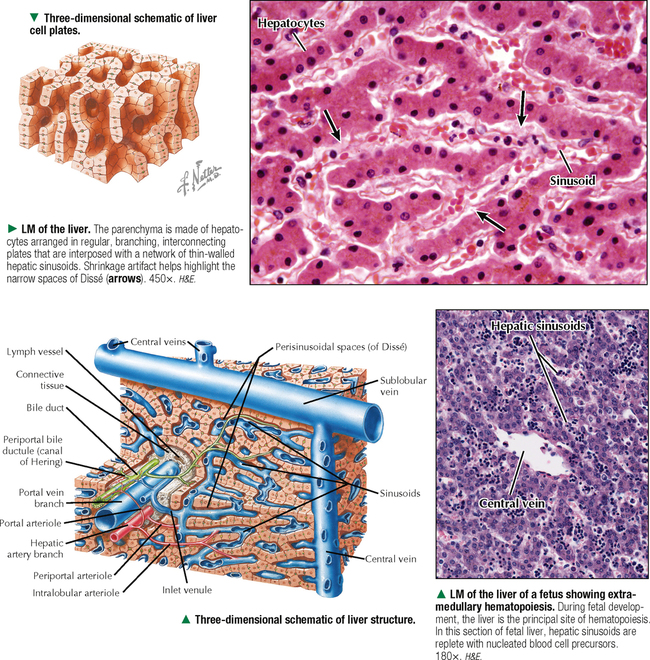
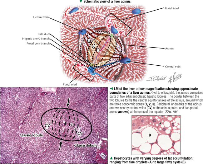
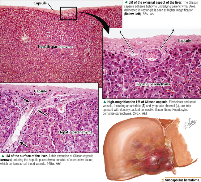
14 LIVER, GALLBLADDER, AND EXOCRINE PANCREAS 14.1. Overview of the Liver 14.2. Classic Hepatic Lobules 14.3. Portal Triads with Blood and Bile Supply 14.4. Histology of the Portal Tract and Central Vein 14.5. Histologic Arrangement of Hepatic Parenchyma 14.6. Structure and Function of the Liver Acinus 14.7. Histology of Glisson Capsule 14.8. Ultrastructure of Hepatocytes 14.9. Ultrastructure and Function of Hepatocytes 14.10. Ultrastructure of Hepatic Sinusoids 14.11. Ultrastructure and Function of Kupffer Cells 14.12. Ultrastructure of the Space of Dissé 14.13. Ultrastructure and Function of Hepatic Stellate Cells 14.14. Histology and Ultrastructure of the Hepatic Biliary Duct System 14.15. Ultrastructure and Function of Bile Canaliculi 14.16. Overview of the Gallbladder 14.17. Histology of the Gallbladder Wall 14.18. Ultrastructure and Function of the Gallbladder Mucosa 14.19. Overview of the Pancreas 14.20. Histology of the Exocrine Pancreas: Ducts 14.21. Histology of the Exocrine Pancreas: Acini 14.22. Ultrastructure of the Exocrine Pancreas 14.23. Development of the Pancreas 14.1 OVERVIEW OF THE LIVER The wedge-shaped liver, the largest and heaviest internal organ (weighs about 1.5 kg in an adult), is essential to life and is the most versatile and vascular organ. It sits just below the diaphragm in the upper right quadrant of the abdominal cavity and is protected completely by the rib cage. It comprises two main lobes of almost equal size—right and left—separated by a falciform ligament and a round ligament (ligamentum teres). Two smaller lobes—caudate and quadrate—are seen on the inferior (visceral) surface but are poorly demarcated. Under a peritoneal serous covering, a thin connective tissue (Glisson) capsule surrounds the lobes. On the visceral surface is the porta hepatis, the gateway for the hepatic ducts, portal vein, hepatic artery, lymphatics, and nerves. The liver arises in the embryo as a diverticulum of foregut endoderm. Parenchymal cells proliferating from this diverticulum pass into mesenchyme of the septum transversum and occupy meshes of a network of capillaries related to vitelline and umbilical vessels. The liver is an accessory gland—both exocrine and endocrine—of the digestive tract and plays a central role in three broad functional categories: metabolism of carbohydrates, proteins, and fats; modification of exogenous substances such as drugs and alcohol; and formation and exocrine secretion of bile, which is critical for fat digestion. CLINICAL POINT Hepatitis—inflammation of the liver—is a serious health risk worldwide. Caused by hepatotropic viruses (A to G), viral hepatitis encompasses a broad array of acute or chronic inflammatory liver disorders. Epstein-Barr virus, herpes simplex virus, and cytomegalovirus may also cause acute hepatitis. Modes of transmission are fecal-oral, food/waterborne, sexual, parenteral, and perinatal. Acute hepatocellular injury, elevated serum aminotransferase and bilirubin levels, and the presence of serum IgM and anti–hepatitis A virus antibody characterize hepatitis A. The histologic hallmark of hepatitis B is the presence of ground-glass hepatocytes caused by accumulation of hepatitis B surface antigen in endoplasmic reticulum. Hepatitis C is the leading cause of death from liver disease in North America. Often transmitted via blood or blood products, it may become chronic, which may lead to cirrhosis and hepatocellular carcinoma. 14.2 CLASSIC HEPATIC LOBULES About 80% of liver tissue in the adult is parenchyma consisting of hepatocytes arranged as a labyrinth of cellular plates. The remaining 20% is stroma, a delicate supportive framework of connective tissue that forms the outer Glisson capsule. At the porta hepatis, this capsule is continuous with the arborization of connective tissue that accompanies the branching pattern of the entering hepatic artery and portal vein and the emerging bile duct. Branches of these three structures are the portal triads. They travel together throughout the liver’s interior and divide repeatedly through 17–20 orders of branches. Their size progressively decreases to the terminal ramifications. Connective tissue in the liver indistinctly divides hepatic parenchyma into classic hepatic lobules, which are the liver’s structural units. In humans, the lobules are poorly defined, the amount of connective tissue between lobules being scanty. In each lobule, a delicate stroma of reticular fibers forms a supportive network for hepatocytes and surrounding sinusoids. CLINICAL POINT Malignant tumors of the liver have high morbidity and mortality rates; they may be classified as primary or metastatic. Hepatocellular carcinoma—the most common primary hepatic neoplasia—usually arises from hepatocytes. Chronic liver disease—most often associated with persistent hepatitis B or hepatitis C infection—is the leading cause. Clinical signs may include hepatomegaly, jaundice, fatigue, and elevated serum levels of certain liver enzymes. More commonly, the liver is involved in metastatic (or secondary) spread of tumors from other sites. In most patients with metastatic liver disease, such malignancies mainly arise from the lung, colon, pancreas, and breast. However, other kinds of tumors (e.g., leukemias, lymphomas, melanomas) may also spread to the liver. Depending on the stage of disease, treatment options vary and include surgical resection, targeted radiation therapy, percutaneous hepatic perfusion of chemotherapeutic agents, and liver transplant surgery. 14.3 PORTAL TRIADS WITH BLOOD AND BILE SUPPLY Each classic hepatic lobule has a prism form of about six sides, about 1 mm in diameter and 2 mm long. Portal triads surrounded by small amounts of interlobular connective tissue sit at corners of each lobule in areas called portal tracts, around which are limiting plates of hepatocytes. Triads also mark peripheral meeting places of adjacent lobules, which look like a mosaic of interlocking tiles. In transverse section, each lobule consists of plates of hepatocytes, one or two cells thick, which are separated by hepatic sinusoids and appear to radiate out from a small central vein. Hepatocyte arrangement resembles that of a sponge, with sinusoids represented by the spaces. Each triad consists of a branch of the bile duct, portal vein, and hepatic artery, which divide into smaller branches. Small lymphatic vessels often accompany them. A unique, unusual feature of the liver is a dual blood supply. The portal vein brings nutrient-rich blood from the gastrointestinal tract—75% of the total blood to the liver; hepatic arteries provide 25% of oxygenated blood. The liver receives about 1.5 L of blood each minute, and at least 20% of its volume is occupied by blood. Terminal branches of portal veins, about 300 μm in diameter, regularly give off inlet venules, which empty into thin-walled, fenestrated hepatic sinusoids that are in intimate contact with hepatocytes. Terminal branches of hepatic arteries, which ramify with portal vein branches, end as arterioles that drain into sinusoids, which thus receive a mixture of arterial and venous blood. Sinusoids converge toward a central vein, also called a terminal hepatic venule, and empty into it in the center of each lobule. A central vein is about 50 μm in diameter. Central veins unite to form sublobular veins, which lead into larger hepatic veins that travel alone and branch repeatedly. Hepatic veins coalesce to join the inferior vena cava, the main drainage route of blood from the liver. Important for understanding lobule organization and hepatocyte function, blood and bile flow through lobules in opposite directions. 14.4 HISTOLOGY OF THE PORTAL TRACT AND CENTRAL VEIN At their smallest branches, the three components of the portal triad are accompanied by small lymphatic vessels. Connective tissue stroma known as the portal tract encloses them all. In transverse section, the hepatic arteriole consists of one to three layers of smooth muscle cells and a relatively small lumen. The portal venule has a larger, often collapsed lumen with a more attenuated wall. The bile ductule is lined by simple cuboidal to columnar epithelium and drains exocrine secretions of hepatocytes from the liver. Biliary passages start with tiny bile canaliculi between hepatocytes. Best seen by electron microscopy, they are small intercellular channels formed by groove-like invaginations of adjacent hepatocytes. As canaliculi approach the periphery of each lobule, they are drained by small ducts, known as canals of Hering, lined by a low simple cuboidal epithelium. These canals drain to larger bile ducts in portal tracts. As the ducts widen, their simple columnar epithelium becomes taller. Typical central veins are thin-walled venules with an attenuated endothelium. They normally lack significant investment of connective tissue stroma. The lumen of each central vein has numerous openings, which allows several hepatic sinusoids to drain freely into them. 14.5 HISTOLOGIC ARRANGEMENT OF HEPATIC PARENCHYMA Three-dimensional reconstructions of liver from serial sections provide insights into the arrangement of hepatic parenchyma and its relationship to vascular and biliary duct systems. The parenchyma consists of an anastomosing network of interconnecting plates, one or two cells thick, which resemble walls of a building with spaces in between. The hepatocytes in each plate can be likened to the building’s bricks, and the hepatic sinusoids appear suspended within the spaces. In humans, one-cell-thick plates are most common in the normal adult liver; two-cell-thick plates occur in the embryo and in adults during regeneration in certain diseases. Via electron microscopy or special light microscopic techniques, narrow fluid-filled perivascular spaces—spaces of Dissé (or perisinusoidal spaces)—can be seen separating the endothelial lining of sinusoids from the hepatocyte surfaces. These spaces allow plasma to flow between sinusoidal lumina and hepatocyte surfaces, which permits rapid exchange of soluble, noncellular substances between blood and parenchyma. In the fetus and in chronic anemias, these spaces are sites of extramedullary hematopoiesis. Hepatic lymph originates in these spaces and eventually drains to small lymphatic vessels in portal tracts. 14.6 STRUCTURE AND FUNCTION OF THE LIVER ACINUS Another concept of liver lobulation is the liver acinus—an oval to diamond-shaped area of hepatic parenchyma defined in relation to blood supply from terminal branches of the portal vein and hepatic artery. It is smaller and harder to see than the classic hepatic lobule, but it is useful functionally and clinically because it is best for describing metabolic and pathologic changes in relation to many diseases. Its short axis runs along the border of two classic hepatic lobules; its long axis is an imaginary line between two central veins closest to the short axis. Hepatocytes in the acinus are arranged in three concentric, elliptical zones around the short axis. Zone 1, most central, is closest to the terminal distributing branches of the portal venule and hepatic arteriole. This zone first receives oxygen, hormones, and nutrients from the bloodstream, and most glycogen and plasma protein synthesis by hepatocytes occurs here. Zone 3 is furthest from the distributing vessels; between zones 1 and 3 is the intermediate zone 2. A gradient of metabolic activity exists for many hepatic enzymes in the three zones. Zone 3 is poorly oxygenated, is the first to show ischemic necrosis and fat accumulation if metabolism is altered, and is the site of most drug and alcohol detoxification. The classic hepatic lobule and liver acinus are not contradictory concepts of lobulation but rather complement each other. CLINICAL POINT A broad panel of laboratory tests—known collectively as liver function tests—is used clinically for diagnosis of liver disorders. They also assess disease severity, prognosis, and treatment outcome. Serum aspartate aminotransferase (AST) and alanine transaminase (ALT)—cytosolic enzymes released into bloodstream in response to hepatocyte injury—are sensitive indicators of liver damage. The highest elevations occur in patients with acute viral hepatitis and toxin-induced hepatic necrosis. The AST-to-ALT ratio may also be useful in distinguishing different causes of hepatotoxicity. A high ratio suggests advanced alcoholic liver disease; lower values are seen in those with viral hepatitis. Whereas chronic disorders, such as cirrhosis, lead to decreased serum levels of albumin (a protein synthesized exclusively in the liver), bile duct obstruction and intrahepatic cholestasis cause elevations in alkaline phosphatase (an enzyme present in the biliary duct system). 14.7 HISTOLOGY OF GLISSON CAPSULE Only gold members can continue reading. Log In or Register to continue Share this: Share on X (Opens in new window) X Share on Facebook (Opens in new window) Facebook Like this:Like Loading… Related Related posts: CARDIOVASCULAR SYSTEM SPECIAL SENSES THE CELL FEMALE REPRODUCTIVE SYSTEM Stay updated, free articles. Join our Telegram channel Join Tags: Netters Essential Histology Jun 18, 2016 | Posted by admin in HISTOLOGY | Comments Off on LIVER, GALLBLADDER, AND EXOCRINE PANCREAS Full access? Get Clinical Tree
14 LIVER, GALLBLADDER, AND EXOCRINE PANCREAS 14.1. Overview of the Liver 14.2. Classic Hepatic Lobules 14.3. Portal Triads with Blood and Bile Supply 14.4. Histology of the Portal Tract and Central Vein 14.5. Histologic Arrangement of Hepatic Parenchyma 14.6. Structure and Function of the Liver Acinus 14.7. Histology of Glisson Capsule 14.8. Ultrastructure of Hepatocytes 14.9. Ultrastructure and Function of Hepatocytes 14.10. Ultrastructure of Hepatic Sinusoids 14.11. Ultrastructure and Function of Kupffer Cells 14.12. Ultrastructure of the Space of Dissé 14.13. Ultrastructure and Function of Hepatic Stellate Cells 14.14. Histology and Ultrastructure of the Hepatic Biliary Duct System 14.15. Ultrastructure and Function of Bile Canaliculi 14.16. Overview of the Gallbladder 14.17. Histology of the Gallbladder Wall 14.18. Ultrastructure and Function of the Gallbladder Mucosa 14.19. Overview of the Pancreas 14.20. Histology of the Exocrine Pancreas: Ducts 14.21. Histology of the Exocrine Pancreas: Acini 14.22. Ultrastructure of the Exocrine Pancreas 14.23. Development of the Pancreas 14.1 OVERVIEW OF THE LIVER The wedge-shaped liver, the largest and heaviest internal organ (weighs about 1.5 kg in an adult), is essential to life and is the most versatile and vascular organ. It sits just below the diaphragm in the upper right quadrant of the abdominal cavity and is protected completely by the rib cage. It comprises two main lobes of almost equal size—right and left—separated by a falciform ligament and a round ligament (ligamentum teres). Two smaller lobes—caudate and quadrate—are seen on the inferior (visceral) surface but are poorly demarcated. Under a peritoneal serous covering, a thin connective tissue (Glisson) capsule surrounds the lobes. On the visceral surface is the porta hepatis, the gateway for the hepatic ducts, portal vein, hepatic artery, lymphatics, and nerves. The liver arises in the embryo as a diverticulum of foregut endoderm. Parenchymal cells proliferating from this diverticulum pass into mesenchyme of the septum transversum and occupy meshes of a network of capillaries related to vitelline and umbilical vessels. The liver is an accessory gland—both exocrine and endocrine—of the digestive tract and plays a central role in three broad functional categories: metabolism of carbohydrates, proteins, and fats; modification of exogenous substances such as drugs and alcohol; and formation and exocrine secretion of bile, which is critical for fat digestion. CLINICAL POINT Hepatitis—inflammation of the liver—is a serious health risk worldwide. Caused by hepatotropic viruses (A to G), viral hepatitis encompasses a broad array of acute or chronic inflammatory liver disorders. Epstein-Barr virus, herpes simplex virus, and cytomegalovirus may also cause acute hepatitis. Modes of transmission are fecal-oral, food/waterborne, sexual, parenteral, and perinatal. Acute hepatocellular injury, elevated serum aminotransferase and bilirubin levels, and the presence of serum IgM and anti–hepatitis A virus antibody characterize hepatitis A. The histologic hallmark of hepatitis B is the presence of ground-glass hepatocytes caused by accumulation of hepatitis B surface antigen in endoplasmic reticulum. Hepatitis C is the leading cause of death from liver disease in North America. Often transmitted via blood or blood products, it may become chronic, which may lead to cirrhosis and hepatocellular carcinoma. 14.2 CLASSIC HEPATIC LOBULES About 80% of liver tissue in the adult is parenchyma consisting of hepatocytes arranged as a labyrinth of cellular plates. The remaining 20% is stroma, a delicate supportive framework of connective tissue that forms the outer Glisson capsule. At the porta hepatis, this capsule is continuous with the arborization of connective tissue that accompanies the branching pattern of the entering hepatic artery and portal vein and the emerging bile duct. Branches of these three structures are the portal triads. They travel together throughout the liver’s interior and divide repeatedly through 17–20 orders of branches. Their size progressively decreases to the terminal ramifications. Connective tissue in the liver indistinctly divides hepatic parenchyma into classic hepatic lobules, which are the liver’s structural units. In humans, the lobules are poorly defined, the amount of connective tissue between lobules being scanty. In each lobule, a delicate stroma of reticular fibers forms a supportive network for hepatocytes and surrounding sinusoids. CLINICAL POINT Malignant tumors of the liver have high morbidity and mortality rates; they may be classified as primary or metastatic. Hepatocellular carcinoma—the most common primary hepatic neoplasia—usually arises from hepatocytes. Chronic liver disease—most often associated with persistent hepatitis B or hepatitis C infection—is the leading cause. Clinical signs may include hepatomegaly, jaundice, fatigue, and elevated serum levels of certain liver enzymes. More commonly, the liver is involved in metastatic (or secondary) spread of tumors from other sites. In most patients with metastatic liver disease, such malignancies mainly arise from the lung, colon, pancreas, and breast. However, other kinds of tumors (e.g., leukemias, lymphomas, melanomas) may also spread to the liver. Depending on the stage of disease, treatment options vary and include surgical resection, targeted radiation therapy, percutaneous hepatic perfusion of chemotherapeutic agents, and liver transplant surgery. 14.3 PORTAL TRIADS WITH BLOOD AND BILE SUPPLY Each classic hepatic lobule has a prism form of about six sides, about 1 mm in diameter and 2 mm long. Portal triads surrounded by small amounts of interlobular connective tissue sit at corners of each lobule in areas called portal tracts, around which are limiting plates of hepatocytes. Triads also mark peripheral meeting places of adjacent lobules, which look like a mosaic of interlocking tiles. In transverse section, each lobule consists of plates of hepatocytes, one or two cells thick, which are separated by hepatic sinusoids and appear to radiate out from a small central vein. Hepatocyte arrangement resembles that of a sponge, with sinusoids represented by the spaces. Each triad consists of a branch of the bile duct, portal vein, and hepatic artery, which divide into smaller branches. Small lymphatic vessels often accompany them. A unique, unusual feature of the liver is a dual blood supply. The portal vein brings nutrient-rich blood from the gastrointestinal tract—75% of the total blood to the liver; hepatic arteries provide 25% of oxygenated blood. The liver receives about 1.5 L of blood each minute, and at least 20% of its volume is occupied by blood. Terminal branches of portal veins, about 300 μm in diameter, regularly give off inlet venules, which empty into thin-walled, fenestrated hepatic sinusoids that are in intimate contact with hepatocytes. Terminal branches of hepatic arteries, which ramify with portal vein branches, end as arterioles that drain into sinusoids, which thus receive a mixture of arterial and venous blood. Sinusoids converge toward a central vein, also called a terminal hepatic venule, and empty into it in the center of each lobule. A central vein is about 50 μm in diameter. Central veins unite to form sublobular veins, which lead into larger hepatic veins that travel alone and branch repeatedly. Hepatic veins coalesce to join the inferior vena cava, the main drainage route of blood from the liver. Important for understanding lobule organization and hepatocyte function, blood and bile flow through lobules in opposite directions. 14.4 HISTOLOGY OF THE PORTAL TRACT AND CENTRAL VEIN At their smallest branches, the three components of the portal triad are accompanied by small lymphatic vessels. Connective tissue stroma known as the portal tract encloses them all. In transverse section, the hepatic arteriole consists of one to three layers of smooth muscle cells and a relatively small lumen. The portal venule has a larger, often collapsed lumen with a more attenuated wall. The bile ductule is lined by simple cuboidal to columnar epithelium and drains exocrine secretions of hepatocytes from the liver. Biliary passages start with tiny bile canaliculi between hepatocytes. Best seen by electron microscopy, they are small intercellular channels formed by groove-like invaginations of adjacent hepatocytes. As canaliculi approach the periphery of each lobule, they are drained by small ducts, known as canals of Hering, lined by a low simple cuboidal epithelium. These canals drain to larger bile ducts in portal tracts. As the ducts widen, their simple columnar epithelium becomes taller. Typical central veins are thin-walled venules with an attenuated endothelium. They normally lack significant investment of connective tissue stroma. The lumen of each central vein has numerous openings, which allows several hepatic sinusoids to drain freely into them. 14.5 HISTOLOGIC ARRANGEMENT OF HEPATIC PARENCHYMA Three-dimensional reconstructions of liver from serial sections provide insights into the arrangement of hepatic parenchyma and its relationship to vascular and biliary duct systems. The parenchyma consists of an anastomosing network of interconnecting plates, one or two cells thick, which resemble walls of a building with spaces in between. The hepatocytes in each plate can be likened to the building’s bricks, and the hepatic sinusoids appear suspended within the spaces. In humans, one-cell-thick plates are most common in the normal adult liver; two-cell-thick plates occur in the embryo and in adults during regeneration in certain diseases. Via electron microscopy or special light microscopic techniques, narrow fluid-filled perivascular spaces—spaces of Dissé (or perisinusoidal spaces)—can be seen separating the endothelial lining of sinusoids from the hepatocyte surfaces. These spaces allow plasma to flow between sinusoidal lumina and hepatocyte surfaces, which permits rapid exchange of soluble, noncellular substances between blood and parenchyma. In the fetus and in chronic anemias, these spaces are sites of extramedullary hematopoiesis. Hepatic lymph originates in these spaces and eventually drains to small lymphatic vessels in portal tracts. 14.6 STRUCTURE AND FUNCTION OF THE LIVER ACINUS Another concept of liver lobulation is the liver acinus—an oval to diamond-shaped area of hepatic parenchyma defined in relation to blood supply from terminal branches of the portal vein and hepatic artery. It is smaller and harder to see than the classic hepatic lobule, but it is useful functionally and clinically because it is best for describing metabolic and pathologic changes in relation to many diseases. Its short axis runs along the border of two classic hepatic lobules; its long axis is an imaginary line between two central veins closest to the short axis. Hepatocytes in the acinus are arranged in three concentric, elliptical zones around the short axis. Zone 1, most central, is closest to the terminal distributing branches of the portal venule and hepatic arteriole. This zone first receives oxygen, hormones, and nutrients from the bloodstream, and most glycogen and plasma protein synthesis by hepatocytes occurs here. Zone 3 is furthest from the distributing vessels; between zones 1 and 3 is the intermediate zone 2. A gradient of metabolic activity exists for many hepatic enzymes in the three zones. Zone 3 is poorly oxygenated, is the first to show ischemic necrosis and fat accumulation if metabolism is altered, and is the site of most drug and alcohol detoxification. The classic hepatic lobule and liver acinus are not contradictory concepts of lobulation but rather complement each other. CLINICAL POINT A broad panel of laboratory tests—known collectively as liver function tests—is used clinically for diagnosis of liver disorders. They also assess disease severity, prognosis, and treatment outcome. Serum aspartate aminotransferase (AST) and alanine transaminase (ALT)—cytosolic enzymes released into bloodstream in response to hepatocyte injury—are sensitive indicators of liver damage. The highest elevations occur in patients with acute viral hepatitis and toxin-induced hepatic necrosis. The AST-to-ALT ratio may also be useful in distinguishing different causes of hepatotoxicity. A high ratio suggests advanced alcoholic liver disease; lower values are seen in those with viral hepatitis. Whereas chronic disorders, such as cirrhosis, lead to decreased serum levels of albumin (a protein synthesized exclusively in the liver), bile duct obstruction and intrahepatic cholestasis cause elevations in alkaline phosphatase (an enzyme present in the biliary duct system). 14.7 HISTOLOGY OF GLISSON CAPSULE Only gold members can continue reading. Log In or Register to continue Share this: Share on X (Opens in new window) X Share on Facebook (Opens in new window) Facebook Like this:Like Loading… Related Related posts: CARDIOVASCULAR SYSTEM SPECIAL SENSES THE CELL FEMALE REPRODUCTIVE SYSTEM Stay updated, free articles. Join our Telegram channel Join Tags: Netters Essential Histology Jun 18, 2016 | Posted by admin in HISTOLOGY | Comments Off on LIVER, GALLBLADDER, AND EXOCRINE PANCREAS Full access? Get Clinical Tree