Acute Hepatitis
This refers to inflammation of the liver with little or no fibrosis and little or no nodular regeneration. There may be minor distortion of lobular architecture. If there is extensive fibrosis with nodular regeneration (and hence distortion of architecture) the condition is called cirrhosis. These diagnoses are made histologically and there may or may not be clinical evidence of previous hepatic disease.
Inflammation with necrosis of liver cells results from:
- Infection, most commonly acute infectious hepatitis A, but also with the viruses of hepatitis B, C and E, infectious mononucleosis, cytomegalovirus (CMV) and yellow fever, and associated with septicaemia and leptospirosis. Amoebic hepatitis is common on a worldwide basis and usually presents as a hepatic abscess or amoeboma.
- Chemical poisons and drugs are less frequent causes of acute hepatitis. Toxic chemicals include carbon tetrachloride, vinyl chloride, and ethylene glycol and similar solvents (glue sniffing). Toxic drugs include alcohol (ethanol and methanol), halothane (after repeated exposures), isoniazid and rifampicin, paracetamol, methotrexate, chlorpromazine and the monoamine oxidase inhibitors.
- Pregnancy (rare).
If the patient recovers this is usually complete, but, rarely, progressive necrosis may affect almost the entire liver (fulminant hepatic failure or acute massive necrosis) causing hepatic coma (p. 149) and death.
Viral Hepatitis
The clinical features of acute hepatitis A, B, C and E are similar, although they differ in severity, time course and progression to chronic liver disease.
Hepatitis A
Hepatitis A (infectious hepatitis) is a single-stranded RNA picornavirus of the enterovirus family which is excreted in the stool towards the end of the incubation period and disappears as the illness develops. Anti-hepatitis A virus immunoglobulin M (IgM) appears at the onset of the illness and indicates recent infection. The disease is endemic but small epidemics may occur in schools or institutions. Spread is usually via the faecal–oral route by food products such as shellfish.
Clinical Presentation
After an incubation period of 2–6 weeks there is gradual onset of influenza-like illness with fever, malaise, anorexia, nausea, vomiting and upper abdominal discomfort associated with tender enlargement of the liver and, less commonly, the spleen. In smokers, there may be a distaste for cigarettes. After 3–4 days the urine becomes characteristically dark and the stools pale – evidence of cholestasis. Symptoms usually become less severe as jaundice appears, although pruritus may develop. Jaundice and symptoms tend to improve after 1–2 weeks and recovery is usually complete, although mild symptoms continue for 3–4 months in a few patients. Recurrent hepatitis A is extremely rare and immunity probably lifelong.
Diagnosis
Diagnosis depends on detecting anti-hepatitis A virus IgM in serum.
Differential Diagnosis
- Obstructive jaundice, either in the early cholestatic phase or in the rare case where cholestatic jaundice persists after other clinical and biochemical evidence of liver cell damage has settled. It is dangerous to diagnose infective hepatitis in patients over 40 years old – a safeguard against misdiagnosing major bile duct obstruction.
- Drug jaundice (p. 152).
- Glandular fever.
- Yellow fever (travellers).
- Acute alcoholic hepatitis may present with enlargement and tenderness of the liver and, sometimes, obstructive jaundice. There are usually other signs of alcoholism.
- Wilson’s disease must not be overlooked (pp. 152 and 205).
Management
If hospitalised, the patient should be isolated. Virus is present in stools for 1–2 weeks before the onset of jaundice and for 1 week after. Symptomatic treatment only is required in the active disease state. No dietary restriction, other than alcohol, is necessary. Liver function tests usually return completely to normal in 1–3 months.
Recovery is the rule in virtually every case. Occasionally jaundice may be prolonged by intrahepatic cholestasis, and corticosteroids can be used to reduce the jaundice rapidly, particularly if associated with pruritus.
Fulminant hepatic failure is rare but has a mortality of about 50%. Management should be in a specialised centre where liver transplantation can be considered.
Prophylaxis
Active immunisation with inactivated virus is recommended for travellers to endemic areas.
Passive immunisation with pooled human immunoglobulin gives partial, short-lived immunity, but is effective immediately after the injection.
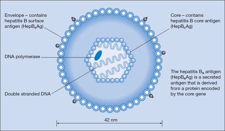
Hepatitis B (Fig. 13.1)
Hepatitis B is a double-stranded DNA hepadnavirus. It is spread by infected blood and serum and also occurs in saliva, semen and vaginal secretions. It is most frequently transmitted by sexual activity, shared needles used by drug addicts, and from mother to child. Health workers and other at-risk groups are now screened and offered vaccination.
Hepatitis B virus causes acute liver disease that ranges from subclinical to fulminant hepatitis, and chronic diseases including chronic hepatitis, cirrhosis and hepatocellular carcinoma.
Clinical Features
After a long incubation period of 6 weeks to 6 months, there is typically a gradual onset of lethargy, anorexia, abdominal discomfort, jaundice and hepatomegaly. It is often asymptomatic in infants. Occasionally immune-mediated extrahepatic manifestations occur with polyarthritis, skin rashes and glomerulonephritis. Cholestatic hepatitis is rare. Mutated viral strains may be associated with fulminant hepatic failure.
Diagnosis
Hepatitis B virus has three different antigens: a surface antigen (HepBsAg), a core antigen (HepBcAg) and an internal component (HepBeAg). HepBsAg appears in the blood about 6 weeks after acute infection and has usually gone by 3 months. HepBeAg occurs at a similar time and denotes high infectivity. HepBcAg is usually found only in the liver. The development of antibodies to HepBsAg usually follows acute infection and indicates immunity. In about 5% of cases antibodies do not appear and HepBsAg persists in the blood (carrier state).
Hepatitis D virus (Delta virus) is an incomplete RNA virus that depends on the hepatitis B virus to replicate. It can cause an aggressive chronic hepatitis in HepBsAg-positive patients. Chronic infection is usually associated with progressive liver disease.
Management
Isolate if hospitalised. In the majority of cases spontaneous recovery occurs and treatment is supportive, as for hepatitis A. Of the estimated 50 million new cases of hepatits B diagnosed worldwide 75% occur in Asia, and 5–10% of adults and up to 90% of children become chronically infected. Prevalence in the UK is < 2%. The carrier state is usually asymptomatic but is associated with chronic hepatitis, which may progress to cirrhosis and hepatocellular carcinoma. Antiviral agents include interferon-α2b, pegylated interferon-α2a, the nucleoside analogue lamivudine and telbivudine, and the nucleotide analogues adefovir and entecavir. Hepatitis B viral DNA becomes undetectable in up to 75% of patients treated for 1 year with nucleoside analogues, but recurrence of viral DNA after treatment withdrawal is common and viral resistance occurs with prolonged use. Pegylated interferon-α2a is more expensive but 1-year seroconversion rates of 30% have been reported.
Immunisation
Immunisation using recombinant HepBsAg is advised for occupational high-risk groups, including health workers, patients with chronic renal failure, intravenous drug abusers, individuals who change sexual partners frequently, patients who receive regular blood products, and family contacts of a case or carrier. Immunisation takes up to 6 months to confer immunity, and booster is recommended after 5 years.
Hepatitis C
Hepatitis C is a single-stranded RNA flavivirus. It predominantly affects intravenous drug abusers and patients who have received multiple blood transfusions. It accounts for 20% of acute hepatitis, 70% of chronic hepatitis, nearly half the cases of end-stage cirrhosis, 60% of primary liver cancer and 30% of liver transplants in the UK.
Clinical Features
Frequently asymptomatic – fewer than 10% of adults become jaundiced. Acute hepatitis following blood transfusion has been virtually eradicated by the introduction of testing of blood products for hepatitis B and C. The incubation period varies from 2 to 26 weeks. Sixty to eighty percent of those acutely infected develop chronic infection, which leads to cirrhosis in around 25% of patients over a 20- to 30-year period. Hepatocellular carcinoma has an annual incidence of around 3% in those with cirrhosis.
Diagnosis
Diagnosis is usually by antibody detection. First- generation enzyme-linked immunosorbent assay (ELISA) using recombinant antigen C100 is relatively non-specific. Newer assays using putative core antigens are more specific, although false positives still occur. The mean time from infection to antibody detection is 12 weeks. Hepatitis C virus RNA determination by PCR (polymerase chain reaction) is used to monitor hepatitis C infection.
Management
Progression to chronic active hepatitis and cirrhosis is much more common in hepatitis C than hepatitis B infection. Combination therapy with pegylated interferon-α and the nucleotide analogue ribavirin achieves a sustained virological response in 40–50% of individuals infected with hepatitis C genotype 1, which is prevalent in Western countries.
Hepatitis E
Hepatitis E virus is an RNA hepevirus that is endemic in the developing world. Transmission is faecal–oral.
Clinical Features
Infection is usually self-limiting, and it does not have a carrier state, but can cause fulminant hepatic failure in pregnant women, with mortality rates of around 20%.
Hepatitis G
Hepatitis G is a positive-stranded RNA flavivirus. There is no evidence at present that it causes acute or chronic liver disease.
Autoimmune Hepatitis
Autoimmune hepatitis is characterised by hepatocellular inflammation and necrosis, which tends to progress to cirrhosis. It may be triggered by acute viral hepatitis and can coexist with chronic viral hepatitis. It has been classified into two types based on the presence of autoantibodies:
Type I autoimmune hepatitis is the commonest type, with a female : male ratio of 8 : 1. It is characterised by the presence of anti-nuclear antibody (50–70%) or anti-smooth muscle cell antibody (50–80%). Anti-mitochondrial antibodies are present in 20% of cases. IgG concentrations and serum aminotransferases are elevated. Liver histology reveals plasma cell infiltrates, liver cell rosettes and piecemeal necrosis. It is associated with HLA B8, DR3 and DR4.
Type II autoimmune hepatitis typically affects girls aged 2–14 years, although 20–30% of cases occur in adults. Onset is often acute, with rapid progression to liver failure. It is characterised by the presence of anti-liver kidney microsome antibodies or anti-liver cytosol type 1 antibodies in the absence of antinuclear or antismooth muscle cell antibodies. It is associated with HLA B14, DR3 and the complement allele C4AQO.
Management
Immunosuppressive therapy improves survival in the majority of cases. Eighty percent of patients respond to steroids, alone or in combination with azathioprine. In patients who fail to respond, orthotopic liver transplantation is the treatment of choice.
Alcoholic Hepatitis
Moderate alcohol consumption is associated with a reduction in mortality compared with either abstinence or heavy drinking (Trials Box 13.1). Drinking in excess of 3 units of alcohol daily may increase mortality, but sensitivity to alcohol varies between individuals (8 g = 1 unit of ethanol = present in a single (25-ml) measure of spirits; a small (125-ml) glass of 12% wine contains 1.5 units). The clinical features of alcoholic liver disease vary from no clinical evidence at all, through nausea, episodes of right abdominal pain associated with tender hepatomegaly, fever and polymorphic leukocytosis, to cirrhosis with portal hypertension and fulminant hepatic failure. Marked jaundice is not always present.
Stay updated, free articles. Join our Telegram channel

Full access? Get Clinical Tree


