Patient Story
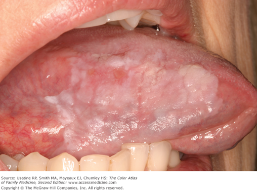
A 57-year-old male smoker presents at the physician’s clinic with a 7-month history of a nonpainful white patch below his tongue. He admits to drinking 2 to 3 beers in the evening and smoking 1 pack of cigarettes per day. Your examination reveals a painless white, thick lesion with fissuring below the tongue (Figure 42-1). A biopsy shows this to be premalignant and the patient is told that he must stop smoking and drinking. He is also referred to an oral surgeon for further evaluation of his dysplasia.
Introduction
The World Health Organization defines leukoplakia as a clinical term used to recognize “white plaques of questionable risk having excluded (other) known diseases or disorders that carry no increased risk for cancer.”1,2 For all types of leukoplakia (see “Clinical Features” below) the risk of malignant transformation is approximately 1%, with a much higher risk associated with leukoplakias manifesting a red and/or highly variable surface texture component.
The term erythroplakia is reserved for a purely red lesion, which is described as a “fiery red patch that cannot be characterized clinically or pathologically as any other definable disease.”1,2 It may be flat or slightly depressed and exhibits a smooth or granular surface texture. The majority of erythroplakias will undergo malignant transformation.
Synonyms
Epidemiology
Etiology and Pathophysiology
- Both leukoplakia and erythroplakia likely represent clinical changes associated with the underlying multistep progression of alterations at the molecular level underlying the development of dysplasia and subsequent carcinoma.
- For all types of leukoplakia, the risk of malignant transformation is approximately 1%, with a much higher risk associated with leukoplakias manifesting a red component.1
- For erythroplakia, the risk of malignant transformation is extremely high, with 85% of cases demonstrating either dysplasia or carcinoma in situ at the time of biopsy.3
Risk Factors
- Smoking and alcohol exposure are the most prominent risk factors for leukoplakia and erythroplakia, and create a synergistic effect when combined.1
- Human papillomavirus (HPV) is a recognized risk factor for oropharyngeal cancer, but its association with leukoplakia and erythroplakia is undetermined.4
- Up to 27% of leukoplakias are idiopathic.4
Diagnosis
- Both leukoplakia and erythroplakia are clinical working diagnoses of exclusion, to be applied when other conditions have been excluded.
- Leukoplakia may be characterized as either homogenous or nonhomogenous.1,2
- Homogenous leukoplakia (Figure 42-1) presents uniformly as a thin surface plaque with possible shallow surface cracks.2
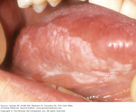
- Nonhomogenous leukoplakia (Figures 42-2 and 42-3) may be further characterized as speckled (white predominant with interspersed red component); nodular (small polypoid outcrops, may be red or white); or verrucous (corrugated or folded surface appearance).2
- Erythroplakia (Figure 42-4) presents as distinct flat or slightly depressed red lesion with a smooth or granular surface texture.1,2