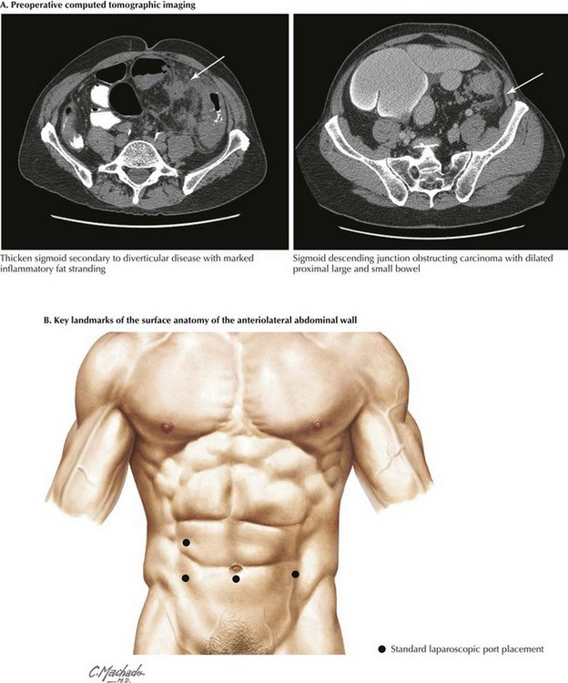
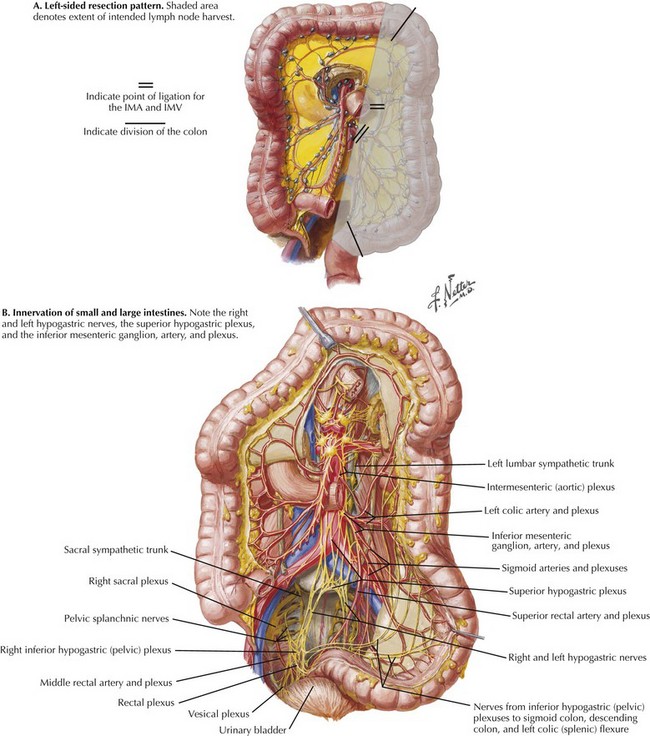
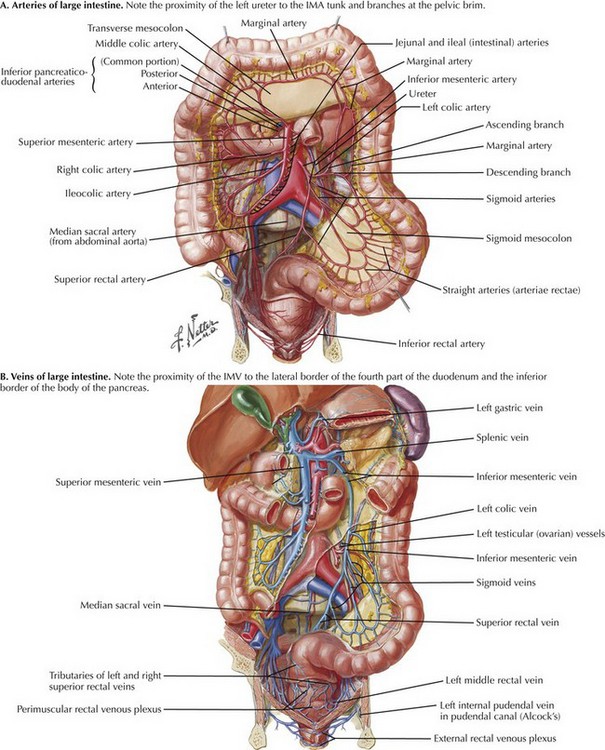
Chapter 22 For colonic carcinoma, computed tomography (CT) of the chest, abdomen, and pelvis provides preoperative staging of the disease. CT includes identification of distant metastases, gross local lymph node involvement, and local invasion of the primary tumor (Fig. 22-1, A). Full colonoscopic evaluation determines the presence of synchronous lesions. For benign disease, CT and colonoscopy may help to identify pathologic features, such as the extent of Crohn disease or ischemia, that may affect approach and extent of dissection. A number of different port configurations have been described; the authors’ preferred approach is shown in Figure 22-1, B. Open left hemicolectomy may be performed through either a midline or a left transverse incision. The anatomy of the vascular supply to the colon is demonstrated in Figure 22-2. Knowledge of these vessels, the autonomic nerves, and the lymphatic drainage, as well as its relationship to the spleen, pancreas, kidney, and ureter, is required for successful completion of the left hemicolectomy (Figs 22-3 and 22-4, A). FIGURE 22–2 Vascular supply to colon.
Left Colectomy

Anatomy for Preoperative Imaging
Surface Anatomy, Incision, and Port Placement
Vascular Anatomy
IMA, Inferior mesenteric artery; IMV, inferior mesenteric vein.
![]()
Stay updated, free articles. Join our Telegram channel

Full access? Get Clinical Tree


Basicmedical Key
Fastest Basicmedical Insight Engine