Large Cell Calcifying Sertoli Cell Tumor
Steven S. Shen, MD, PhD
Jae Y. Ro, MD, PhD
Key Facts
Terminology
Variant of Sertoli cell tumor with large epithelioid cells and peculiar calcifications, often associated with clinical syndromes
Clinical Issues
Extremely rare; only ~ 50 cases have been reported
Range: 1.5-48 years (average: 16 years)
Sporadic (60%) or as a component of Carney and Peutz-Jeghers syndromes
Macroscopic Features
Well-circumscribed white-tan or yellow mass associated with granular or gritty calcification
Usually smaller than 4 cm
Microscopic Pathology
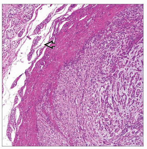
Common growth patterns include nests, cords or trabeculae, or solid tubules
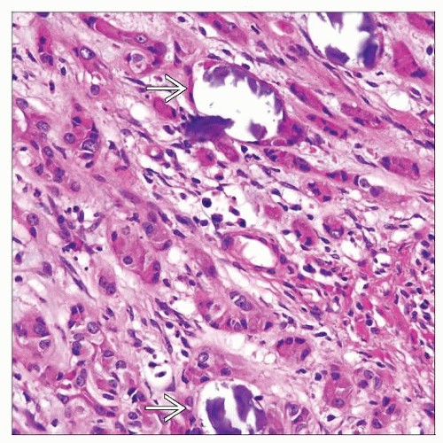
Large polygonal cells with abundant eosinophilic, “ground-glass,” or finely granular cytoplasm
Tumor cells: Vesicular nuclei & prominent nucleoli
Fibrous or myxohyaline stroma with marked calcifications
May have marked neutrophilic infiltration
Some have intratubular growth
Ancillary Tests
Positive for vimentin, inhibin-α, NSE, S100, desmin, actin-sm
Negative for cytokeratin (can be focally positive), α-fetoprotein, HCG, PLAP, Podoplanin, and Oct3/4
TERMINOLOGY
Abbreviations
Large cell calcifying Sertoli cell tumor (LCCSCT)
Definitions
Variant of Sertoli cell tumor with large epithelioid cells and peculiar calcifications, often associated with clinical syndromes
CLINICAL ISSUES
Epidemiology
Incidence
Extremely rare, < 60 have been reported
Age
Range: 1.5-48 years (average: 16 years)
Presentation
Slowly enlarging, painless testicular mass
Sporadic (60%) or as a component of Carney and Peutz-Jeghers syndromes
Some patients may have gynecomastia or precocious puberty
Acromegaly or gigantism may be seen due to associated pituitary adenoma
Association with cardiac myxoma or sudden death
May be associated with adrenocortical hyperplasia, hypercortisolemia, or testicular Leydig cell tumor
High frequency of bilaterality (40%) and multifocality (60%)
Intratubular LCCSCT is a variant of LCCSCT occurring in boys with Peutz-Jeghers syndrome; often estrogenic, bilateral and multifocal; always benign
Malignant form: Mean age: 39 years; more often unilateral and solitary compared to benign tumors
Treatment
Surgical approaches
Orchiectomy is usually curative; long-term followup is necessary
Prognosis
Excellent, but some (20%) may have malignant behavior
IMAGE FINDINGS
Radiographic Findings
Ultrasound may detect testicular mass with brightly echogenic area (calcification)
MACROSCOPIC FEATURES


