Proliferation of atypical keratinocytes typically confined to basilar 1/3 of epidermis
Overlying parakeratosis and basilar budding usually seen
• Solar lentigo (lentigo senilis)
Elongated, hyperpigmented rete ridges
Lacks keratinocyte atypia/pleomorphism
• Seborrheic keratosis
More prominent epidermal acanthosis with pseudohorn cysts
Large Cell Acanthoma LCA shows an intraepidermal proliferation of enlarged keratinocytes in a disorganized pattern. There is no basilar budding or overlying parakeratosis typical of an actinic keratosis. Note the prominent solar elastosis in dermis.
Large Cell Acanthoma at High Magnification Higher power of LCA shows enlarged keratinocytes involving the mid to upper layers of epidermis with overlying basket-weave orthokeratosis. The dermis shows solar elastosis and mild chronic inflammatory infiltrate .
Large Cell Acanthoma at Low Magnification Another example of LCA shows an intraepidermal proliferation of enlarged squamous cells . There is a lack of basilar budding or overlying parakeratosis typical of actinic keratosis.
Large Cell Acanthoma at High Magnification The cells show nuclear enlargement, hyperchromasia, and focally prominent nucleoli , but no mitotic figures are identified.
TERMINOLOGY
Abbreviations
• Large cell acanthoma (LCA)
Definitions
• Proliferation of enlarged and mildly atypical keratinocytes in sun-damaged skin without diagnostic features of actinic keratosis (AK)
Has been variously considered to be related to or variant of solar lentigo, seborrheic keratosis (stucco keratosis) (SK), or AK
ETIOLOGY/PATHOGENESIS
Environmental Exposure
• Most cases associated with UV radiation (chronic solar damage)
Infectious Agents
• HPV types have been detected in some studies
CLINICAL ISSUES
Epidemiology
• Incidence
Uncommon lesions but may be underreported
• Age
Typically occur in older adults (mean: 75 yr)
Only gold members can continue reading. Log In or Register to continue


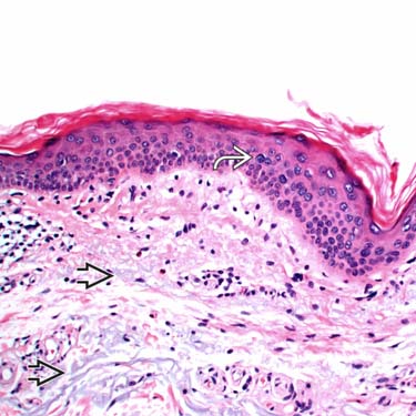
 in a disorganized pattern. There is no basilar budding or overlying parakeratosis typical of an actinic keratosis. Note the prominent solar elastosis
  in dermis.
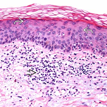
 involving the mid to upper layers of epidermis with overlying basket-weave orthokeratosis. The dermis shows solar elastosis and mild chronic inflammatory infiltrate
  .
.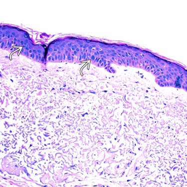
 . There is a lack of basilar budding or overlying parakeratosis typical of actinic keratosis.
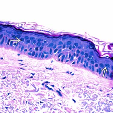
 , but no mitotic figures are identified.


