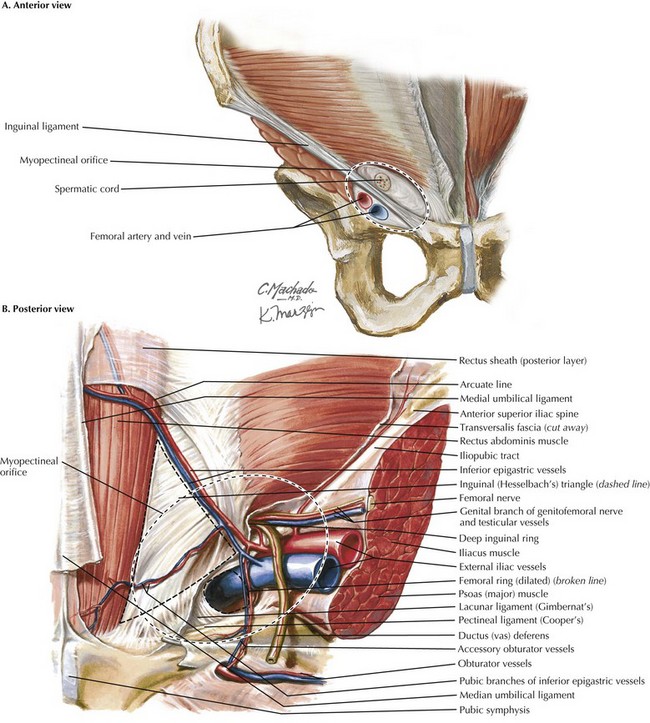
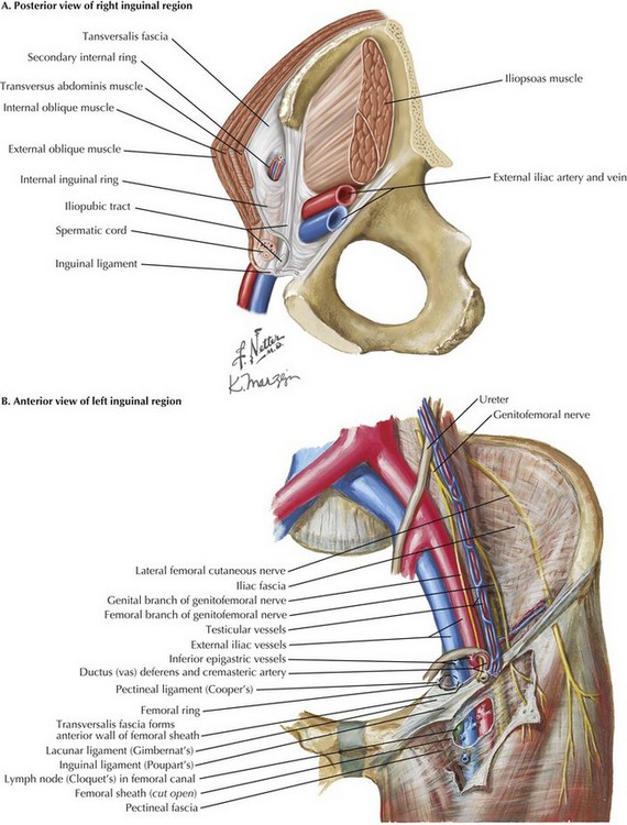
Laparoscopic Inguinal Hernia Repair

Chapter 29


Laparoscopic Inguinal Hernia Repair


Introduction


Inguinal hernias form because of a defect in the myopectineal orifice that allows intraabdominal contents to protrude into the groin. Accounting for approximately 800,000 cases annually in the United States alone, inguinal hernia repair is one of the most common operations performed by general surgeons. Open anterior surgical repair with mesh prosthesis was the technique of choice until the early 1990s, when the introduction of laparoscopy revolutionized inguinal hernia repair. Benefits of the laparoscopic technique include lower incidence of chronic pain and faster return to work. The laparoscopic approach also affords significant advantages for patients with bilateral hernias, recurrent hernias previously repaired by an anterior approach, and femoral hernias. Regardless of the approach, an in-depth knowledge of groin anatomy is essential to achieve a durable repair.


Laparoscopic Approaches


The two most common laparoscopic hernia repair approaches are the transabdominal preperitoneal (TAPP) and the totally extraperitoneal (TEP). In the TAPP technique the abdominal cavity is entered and a transverse incision is made in the peritoneum, starting at the medial umbilical ligament and continuing out laterally just short of the anterior superior iliac spine (ASIS). The peritoneum is peeled down from the transversalis fascia to expose the entire myopectineal orifice and create a “pocket.” Mesh is then placed into this pocket in the preperitoneal position and secured with tacks and/or glue. The peritoneum is then reclosed with suture or tacks, thus excluding the mesh from the intraabdominal contents to prevent bowel adhesions and minimize the risk of intestines being “trapped” in the preperitoneal space.


The TEP approach differs by avoiding entry into the abdominal cavity. Instead, balloon dissection creates a pocket for the mesh between the rectus abdominis muscle and the transversalis fascia.


No significant difference has been found between TAPP and TEP with regard to length of surgery, return to normal activity, or rate of recurrence. Some studies suggest a higher incidence of port-site hernias and visceral injuries with TAPP, whereas more conversions may occur with TEP. Ultimately, surgeons should choose the technique they are most comfortable with to obtain the best outcomes.


Regardless of the approach taken, the goal of laparoscopic herniorrhaphy remains a durable repair. In contrast to the open repair, the failures of the laparoscopic repair occur at the inferior border as the viscera “sneaks in” underneath the inferior edge of the mesh. As a result, sufficient dissection of the pocket along the inferior border is paramount to reduce recurrence. In addition, the authors often use fibrin glue to fixate the inferior edge of the mesh.


Key Anatomic Concepts for Laparoscopic Repair


Myopectineal Orifice


The myopectineal orifice is one of the most important anatomic features of the groin anatomy (Fig. 29-1, dashed ovals). All hernias of the groin originate from this single zone of weakness, which is covered only by transversalis fascia and peritoneum. Bisected by the inguinal ligament, the myopectineal orifice comprises the inguinal canal superiorly and the femoral canal inferiorly. The inferior border consists of the superior pubic ramus and the pectineal (Cooper’s) ligament. Medially, the myopectineal orifice is bordered by the rectus abdominis muscle and the inguinal falx (conjoined tendon). The conjoined (conjoint) tendon (fusion of internal oblique muscle and transversalis fascia) is also the superior border of the orifice. Laterally, the boundaries consist of the iliopsoas muscle and lateral border of the femoral sheath.


Inguinal Ligament versus Ileopubic Tract


Although a key anatomic landmark for open (anterior) inguinal hernia repair, the inguinal ligament is not seen in the laparoscopic (posterior) repair because it is an anterior lamina structure (Fig, 29-2, A). The inguinal (Poupart’s) ligament is the inferior edge of the external oblique aponeurosis, extending from the ASIS to the pubic tubercle, turning posteriorly to form the “shelving edge.” This shelving edge is used to secure the inferior border of the mesh in an open inguinal hernia repair (see Chapter 28). The iliopubic tract is the continuation of the transversus abdominis aponeurosis and fascia. It is located posterior to the inguinal ligament, extends from the pubic tubercle medially, and passes over the femoral vessels to insert on the ASIS laterally. This posterior lamina structure is an important landmark in laparoscopic inguinal hernia repair; lateral to the internal ring, no tacks should be placed below the iliopubic track because of the risk of injury to the lateral femoral cutaneous, genitofemoral, and femoral nerves.


Pectineal Ligament


The pectineal (Cooper’s) ligament refers to the periosteum found along the superior ramus of the pubic bone, posterior to the iliopubic tract (Fig. 29-2, B). The pectineal ligament is an extension of the lacunar (Gimbernat’s) ligament, which connects the inguinal ligament to Cooper’s ligament near their insertion site at the pubic tubercle. Cooper’s ligament is frequently used for medial fixation of the mesh in a laparoscopic hernia repair.

Only gold members can continue reading. Log In or Register to continue

Stay updated, free articles. Join our Telegram channel

Aug 12, 2016 | Posted by in ANATOMY | Comments Off on Laparoscopic Inguinal Hernia Repair

Full access? Get Clinical Tree

Get Clinical Tree app for offline access