• Defined as clonal proliferation of Langerhans cells
Clinical Issues
• Newborns and infants usually present with limited skin or bone lesions
• Erythematous, crusted, vesiculopustular rash
• Common sites include skin, bone and bone marrow, and lung
• M:F = 2:1
• Eosinophilic granuloma is restricted to bone
• Hand-Schüller-Christian disease has multiple organ involvement and diabetes insipidus
• Letterer-Siwe disease is aggressive with multisystem involvement
Microscopic
• Langerhans cells infiltrate dermis and subcutis and often extend throughout epidermis to stratum corneum
• Langerhans cells show
Abundant eosinophilic show cytoplasm
Coffee bean-shaped nucleus
• Large numbers of eosinophils and other inflammatory cells typically present
• Mitoses common, but usually not atypical
Ancillary Tests
• CD1a and S100 immunostaining
• Birbeck granules seen on EM
Top Differential Diagnoses
• Granulomatous infiltrates
• Rosai-Dorfman disease
• Indeterminate cell histiocytosis
• Hodgkin and non-Hodgkin lymphoma
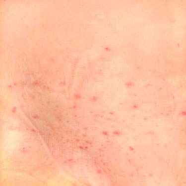
Clinical Photograph of Langerhans Cell Histiocytosis This example of Langerhans cell histiocytosis (LCH) of the skin presented as a seborrheic-like erythematous rash with petechiae on the abdomen of a 23-month-old child. (Courtesy S. Vanderhooft, MD.)
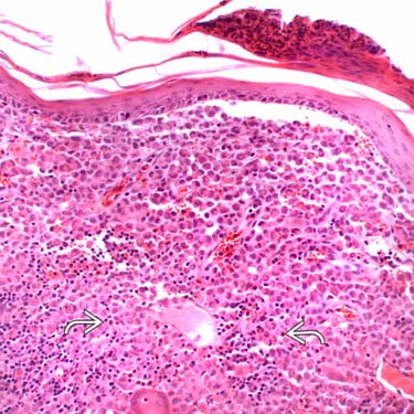
Dermal Involvement by Langerhans Cell Histiocytosis This example of LCH shows dense collections of histiocytic cells in the superficial dermis, with overlying epidermal atrophy and serum crust containing neutrophils. Scattered dermal eosinophils are also seen .
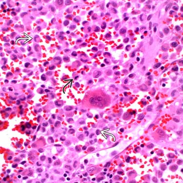
Higher Magnification of Langerhans Cell Histiocytosis Higher magnification shows the characteristic cytologic features, including folded or coffee bean-shaped nuclei , and abundant eosinophilic-staining cytoplasm. This field also shows a mitosis and a few large multinucleated cells, which may seen in some cases.
CD1a Immunohistochemistry in Langerhans Cell Histiocytosis The Langerhans cells show positive cytoplasmic and membranous staining for CD1a.
TERMINOLOGY
Abbreviations
• Langerhans cell histiocytosis (LCH)
Synonyms
• Histiocytosis X
• Eosinophilic granuloma
Definitions
• Clonal proliferation of Langerhans cells
CLINICAL ISSUES
Epidemiology
• Age
First 3 decades of life; rarely occurs in older adults
• Sex
M:F = 2:1
Site
• Skin
Erythematous, crusted, vesiculopustular rash, or salmon-colored macular-papular rash
• Bone and bone marrow
Skull, pelvis, long bones, vertebrae
• CNS
Primary in dura, leptomeninges, or parenchyma
May be secondary to skull or vertebral involvement
• Other organs that may be affected include lung, lymph nodes, liver, thymus, and GI tract
Presentation
• Newborns and infants usually present with limited skin or bone lesions
• Generalized disease is more common in young children
• Rash may precede systemic findings by several months
• Eosinophilic granuloma
Single or multiple lesions restricted to bone
• Hand-Schüller-Christian disease
Multiple organ involvement
Only gold members can continue reading. Log In or Register to continue


 .
.
 , and abundant eosinophilic-staining cytoplasm. This field also shows a mitosis
  and a few large multinucleated cells, which may seen in some cases.


