Kaposi Sarcoma
Thomas Mentzel, MD
Key Facts
Terminology
Locally aggressive endothelial neoplasm associated with human herpes virus 8
Clinical Issues
Most typical site of involvement is skin
Mucosal membranes, lymph nodes, and visceral organs may be affected
4 main different clinical and epidemiologic forms are recognized
Classical indolent form
Endemic African form
Iatrogenic form
AIDS-associated form
Prognosis depends on epidemiological-clinical type
Macroscopic Features
Skin lesions range in size from very small to several centimeters
Microscopic Pathology
Histologic features of all forms of KS do not differ
KS shows different stages of disease
Patch stage of KS
Increased vascular spaces in reticular dermis
Scattered lymphocytes and plasma cells
Plaque stage of KS
More extensive vascular proliferation
Nodular stage of KS
Well-circumscribed, cellular nodules
Intersecting cellular fascicles of spindled tumor cells
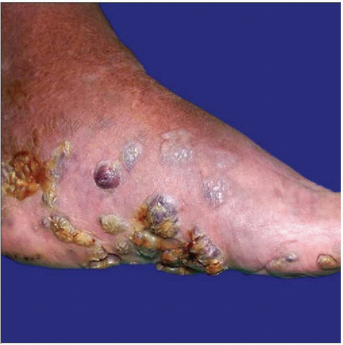 Clinical photograph shows a case of classical Kaposi sarcoma arising in an elderly man who presented with multiple nodular lesions. |
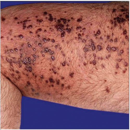 Clinical photograph shows an HIV-positive patient who developed multiple small lesions on the trunk and lower extremities. |
TERMINOLOGY
Abbreviations
Kaposi sarcoma (KS)
Definitions
Locally aggressive endothelial neoplasm associated with human herpes virus 8 (HHV8)
ETIOLOGY/PATHOGENESIS
Infectious Agents
Associated with HHV8
HHV8 is found in all forms of disease
HHV8 is detected in peripheral blood
CLINICAL ISSUES
Site
Most typical site of involvement is skin
Mucosal membranes, lymph nodes, and visceral organs may be affected
Natural History
4 main different clinical and epidemiologic forms are recognized
Classical indolent form
Occur predominantly in elderly men of Mediterranean/East European descent
Purplish, reddish-blue, dark brown plaques and nodules
Usually in distal extremities
Endemic African form
Occurs in middle-aged adults and children in equatorial Africa
Patients are not infected by HIV
Iatrogenic form
Occurs in patients treated by immunosuppressive agents
AIDS-associated form
Occurs in patients infected by HIV-1
Most aggressive form
Lesions are seen on face, genitals, lower extremities
Mucosal membranes, lymph nodes, and visceral organs are frequently involved
Treatment
Options, risks, complications
Chemo- &/or radiotherapy
Cryotherapy may be useful
Surgical approaches
Surgical treatment of single lesions only
Prognosis
Classical indolent form
Indolent clinical course
Lymph node and visceral organ involvement occurs only infrequently
Endemic African form
Protracted clinical course
Lymphadenopathic form is progressive and highly lethal
Iatrogenic form
May resolve entirely after withdrawal of immunosuppressive treatment
AIDS-associated form
Most aggressive type of KS
Prognosis depends on epidemiological-clinical type of KS
Prognosis is strongly related to stage of disease
Prognosis is strongly related to additional infectious diseases
MACROSCOPIC FEATURES
MICROSCOPIC PATHOLOGY
Histologic Features
Histologic features of all forms of KS do not differ
KS shows different stages of disease
Patch stage of KS
Increased vascular spaces in reticular dermis
Papillary dermis is not involved in early stages
Vascular spaces dissect collagen bundles
Perivascular and periadnexal growth of vascular spaces
Vascular spaces are lined by flat, uniform endothelial cells
Scattered lymphocytes and plasma cells
Extravasated erythrocytes and hemosiderin deposits
Plaque stage of KS
More extensive vascular proliferation
Denser inflammatory infiltrate
Hyaline globules representing destroyed erythrocytes may be found
Nodular stage of KS
Well-circumscribed, cellular nodules
Intersecting cellular fascicles of spindled tumor cells
Slit- and sieve-like spaces containing erythrocytes
Mild cytologic atypia
Numerous mitoses
Some patients develop lymphangiomatous lesions &/or hemangiomatous lesions
Cytologic Features
Bland flat and spindled endothelial tumor cells
DIFFERENTIAL DIAGNOSIS
Hobnail Hemangioma
Solitary vascular lesions
Biphasic growth
Dilated vessels in superficial parts, narrow vascular spaces in deeper parts of dermis
Hobnail endothelial cells
HHV8(-)
Capillary Hemangioma
Different clinical findings
Lobular growth of narrow capillaries
HHV8(-)
Lymphangioma
Common pediatric lesions
Rather well-circumscribed lesions
Dilated vascular spaces
Usually no inflammatory infiltrate
HHV8(-)
Progressive Lymphangioma (Benign Lymphangioendothelioma)
Slowly growing, solitary, plaque-like lesions
No spindled tumor cells
No prominent inflammatory infiltrate
HHV8(-)
Spindle Cell Hemangioma
Combination of KS-like features with cavernous hemangioma-like features
Stay updated, free articles. Join our Telegram channel

Full access? Get Clinical Tree


