Iron1


Iron1


Marianne Wessling-Resnick


HISTORICAL PERSPECTIVE

As early as the sixteenth and seventeenth centuries, chlorosis (iron deficiency anemia) was reported as a medical condition that could be treated with iron supplementation, but it was not until the turn of the twentieth century that our understanding of the essential nature of iron for heme synthesis slowly emerged (1). Since that time, the pace of discovery in the field of iron metabolism has accelerated to the current explosion in molecular information in the twenty-first century (2). This chapter highlights the most current concepts in iron homeostasis.


CHEMISTRY AND IMPORTANCE OF IRON

Iron exists in one of two oxidation states: the ferrous form (Fe2+) or the ferric form (Fe3+). This chemical property results in iron’s catalytic role in a multitude of redox reactions necessary to support basic metabolic functions for life. In fact, iron’s central role in oxygen and energy metabolism underscores the biologic significance of this element and helps to explain why it is one of the best-studied metals in nutrition and health. These same catalytic properties of iron also confer its well-known toxicity resulting from Fenton chemistry, a reaction that generates free radicals including superoxide. Thus, iron is an essential nutrient as well as a powerful toxicant, and it is important to understand how both features are kept in balance.

Total iron body content is estimated to be 3.8 g in men and 2.3 g in women. Most body iron is found as heme iron (Fig. 10.1). Heme iron is the essential constituent for oxygen transport in hemoglobin, oxygen storage in myoglobin, and electron transport for cytochrome function in aerobic respiration, and it is even necessary for signal transduction as a cofactor for nitric oxide synthase and guanylyl cyclase. The second largest pool of iron is found in its storage form ferritin (also hemosiderin). Ferritin is a large assembly of 24 protein subunits that form a large sphere around a mineralized ferric core of several thousands of iron atoms (3). In times of demand, iron is liberated from ferritin to fulfill essential functions in oxygen transport and energy metabolism. Damage generated by reactive oxygen species arising from redox-reactive free iron is prevented by its storage in ferritin.

In similar fashion, newly absorbed iron is bound by transferrin, thereby limiting its toxic effects as it is transported in serum. Transferrin-bound iron is destined to be taken up by the transferrin receptor in peripheral tissues for storage or utilization. Transferrin has two binding sites for one iron atom each. Under normal circumstances, 30% to 40% of these iron-binding sites are filled with approximately 4 mg of total body iron. Circulating transferrinbound iron represents a highly dynamic storage pool that can be drawn from to fulfill immediate demands. Hence the saturation state of serum transferrin plays a key role in the regulation of iron metabolism and is one of the indices used clinically to evaluate iron status.


Fig. 10.1. Structure of heme.

Iron absorption, utilization, and storage are finely tuned to maintain the metal’s homeostasis. Unlike the situation with other essential elements, iron metabolism is subject to a high degree of conservation. Rather than eliminate excess iron that is not immediately required, iron is stored in ferritin for times of need, as described earlier. The nature of iron homeostasis reflects the metal’s key chemical role in oxygen and energy metabolism, processes that are necessary for life and must therefore rely on a substantial reservoir of iron to support the ultimate demands of human physiology. Approximately 20 to 25 mg of iron is turned over daily with the erythrophagocytosis of senescent red blood cells, and iron that is released from heme is captured for reutilization in the production of new erythrocytes. Small amounts of iron are lost in feces (˜0.6 mg/day), urine (<0.1 mg/day), and sweat (<0.3 mg/day). Menstruating women suffer an average blood loss of approximately 40 mL/cycle or 0.4 to 0.5 mg/day. Most losses are offset by the amount of iron provided in the diet, however, pathologic conditions associated with excessive blood loss such as hookworm infection or bleeding ulcers can result in greater iron demands. A key feature of iron homeostasis is that the body’s iron status is maintained at the level of dietary iron absorption to prevent toxic accumulation while adequate amounts are provided to offset losses. When iron is depleted from the body, dietary iron absorption increases to meet the demand for iron, although no known regulated pathway exists for excretion of excess iron.


DIETARY SOURCES

Iron is absorbed from the diet as either heme or nonheme iron (see the following website: http://ods.od.nih .gov/factsheets/Iron-HealthProfessional). Heme iron is typically derived from hemoglobin or myoglobin and is contained in foods such as red meats, fish, and poultry. Various sources of nonheme iron, such as plant foods, are also available. This form of iron is also added to enrich and fortify foods such as cereals. Although nonheme iron is the predominant form in the diet, heme iron is more bioavailable (4). Approximately 15% to 35% of heme iron will be assimilated, compared with 2% to 20% nonheme iron absorption. As discussed earlier, the levels of stored iron in the body influence the extent of absorption. Under low-iron conditions, dietary absorption is promoted, whereas high-iron conditions reduce absorption. Many other dietary and endogenous factors can influence iron uptake (Table 10.1). For example, ascorbate can help to reduce ferric iron to the more bioavailable form of ferrous iron (5, 6). Polyphenols and phytates can interfere with nonheme iron uptake (6). Calcium blocks uptake of both heme and nonheme iron (7), whereas other metals can inhibit nonheme iron absorption by sharing the same pathway for absorption (4). In particular, not only is lead a competitive inhibitor for uptake, but also it can disrupt steps of iron metabolism required for heme synthesis (8). Because low iron status enhances metal absorption, lead poisoning is often associated with iron deficiency in children (9).


TABLE 10.1 FACTORS INFLUENCING IRON ABSORPTION


NUTRIENTS


ENDOGENOUS FACTORS


Enhancers


Ascorbic acid (vitamin C)


Enhanced erythropoiesis due to


Fructose


Hypoxia


Citric acid


Hemorrhage


Dietary protein


Hemolysis


Lysine


Androgens


Histidine


Cobalt


Cysteine


Low iron stores


Methionine


Inhibitors


Oxalic acid


Infection/inflammation


Tannins


Lack of stomach acid


Phytate


High iron stores


Polyphenols


Carbonate


Phosphate


Fiber


Other metal ions


Adapted from Linder M. Nutritional Biochemistry and Metabolism with Clinical Applications. New York: Elsevier, 1985, with permission.


RECOMMENDED DIETARY ALLOWANCES

The recommended dietary allowances (RDAs) set for iron by the Institute of Medicine of the National Academy of Sciences are listed in Table 10.2. Iron is considered a micronutrient: adult men require 8 mg iron/day, and during their reproductive years, girls and women require 18 mg iron/day. The typical North American diet of 12 to 18 mg iron/day should be adequate to fulfill these needs, but the requirement for iron markedly increases to 27 mg/day during pregnancy, and iron supplements are often needed to match this high demand. Infants are born with a 4- to 6-month iron supply, and an RDA has not been established for this early age. An adequate intake (AI) of 0.27 mg/day is recommended, however. Beyond this age, iron content in milk cannot entirely meet the needs of the developing child, and food sources are
required to meet RDAs (7 mg/day at age 1 to 3 years and 10 mg/day at age 4 to 8 years). Iron toxicity can also pose a risk for children, and it commonly results from ingestion of excess iron supplements. Death occurs at levels of 200 to 300 mg/kg. The tolerable upper limits (UL) for iron intake are listed in Table 10.2.


TABLE 10.2 RECOMMENDED DIETARY ALLOWANCES AND TOLERABLE UPPER INTAKE LEVELS FOR IRON


RECOMMENDED DIETARY ALLOWANCES


TOLERABLE UPPER INTAKE LEVELS


AGE (y)


MALES (mg/d)


FEMALES (mg/d)


AGE (y)


MALES (mg/d)


FEMALES (mg/d)


Infants


0.58-1.0


11


11


0.58-1.0


40


40


Children


1-3


7


7


1-13


40


40


4-8


10


10


Adolescents


9-13


8


8


14-18


45


45


14-18


11


15


Adults


19-50


8


18


19+


45


45


51+


8


8


Pregnancy


14-18


27


14-18


45


19-50


27


19+


45


Lactation


14-18


10


14-18


45


19-50


9


19+


45


From Food and Nutrition Board, Institute of Medicine. Dietary Reference Intakes for Vitamin A, Vitamin K, Arsenic, Boron, Chromium, Copper, Iodine, Iron, Manganese, Molybdenum, Nickel, Silicon, Vanadium, and Zinc. Washington, DC: National Academy Press, 2001.


IRON METABOLISM AND ITS REGULATION

The field of iron biology has made rapid advances since 2000. Many of the proteins involved in iron transport and homeostatic regulation have been identified, and their physiologic roles have been uncovered. Perhaps the most exciting breakthrough came with the discovery of iron regulatory hormone, hepcidin. Features of hepcidin regulation of iron metabolism have been compared to insulin action in glucose metabolism, thus creating a new field of iron endocrinology (10). How hepcidin regulates systemic iron homeostasis is a major focus of current investigation. At the cellular level, molecular insights into the regulation of iron-binding proteins have yielded information about regulation of iron transport, utilization, and storage that have important clinical considerations. Finally, transcriptional and posttranscriptional networks that may be activated by hepcidin-induced signaling are beginning to emerge and provide clues into the relationships between iron and inflammation.


Intestinal Iron Absorption

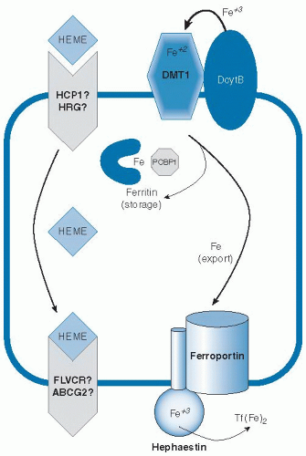
Because body iron status is precisely tuned by the absorption of dietary iron, it is important to understand the mechanisms involved in this process and the multiple layers that regulate the flux of iron into the system (Fig. 10.2). Because the body does not eliminate excess iron, dysregulation of intestinal iron absorption causing assimilation of too much iron will result in iron overload. Conversely, if sufficient iron is not absorbed to make up for small daily losses, the risk for iron deficiency increases. Iron is absorbed as nonheme or heme forms. Heme iron is absorbed more effectively (11), and this process does not appear to be subject to the same regulatory mechanisms as nonheme iron uptake (12).

These findings indicate that heme and nonheme iron are taken up by independent mechanisms. A putative heme transporter called heme carrier protein-1 (HCP1)
was identified (13), but questions about its true function arose when a role in folate transport was determined for the same factor (14). HCP1 may possibly be a low-affinity transporter for heme, but its physiologic relevance remains to be better established. A different molecule, heme-responsive gene-1 or HRG1, has been identified as a heme transporter in Caenorhabditis elegans (15). Although a similar gene is present in humans, its activity has yet to be defined. Heme oxygenase may release iron from heme entering the intestinal absorptive enterocyte to join the pool of newly absorbed nonheme iron entering the cell (16). Alternatively, intact heme may be released across the basolateral surface. Feline leukemia virus C receptor (FLVCR) has been identified to function as a heme exporter in erythroid cells (17), and a second possible efflux pathway involving the ABC transporter ABCG2 (also known as breast cancer regulated protein, BCRP) has been suggested (18), but their possible roles in heme assimilation by the intestine have yet to be fully explored.


Fig. 10.2. Intestinal iron (Fe) absorption. ABCG2, ABC transporter; DcytB, duodenal cytochrome B; DMT1, divalent metal transporter-1; FLVCR, feline leukemia virus C receptor; HCP1, heme carrier protein-1; HRG, heme-responsive gene; PCBP1, poly(rC)-binding protein-1; Tf(Fe)2, diferric transferrin.

Uptake of nonheme iron by enterocytes is better understood. Although less effectively absorbed, nonheme iron is present in a greater range of foods, most typically in the ferric (Fe3+) form. Reduction to Fe2+ is the first step in intestinal nonheme iron assimilation and is mediated by brush-border ferrireductase activity. An enzyme called duodenal cytochrome B (DcytB) has been implicated in this process (19). Although it does not appear to be an essential gene (20), DcytB is highly regulated in response to iron status in animals and humans (21, 22, 23), and a promoter polymorphism observed in the human population appears to modify serum ferritin levels in HFE-associated hereditary hemochromatosis (24). After reduction by DctyB or another brush-border ferrireductase, uptake of the ferrous (Fe2+) form of iron is mediated by divalent metal transporter-1 (DMT1) (25, 26, 27). The low pH of the intestinal lumen is important for these initial steps because DMT1 is a protoncoupled transporter, and acidification is therefore necessary for its optimal activity (26). Like DcytB, DMT1 is also highly regulated by iron status. The small intestine expresses four transcripts of DMT1, and mRNA levels are regulated both transcriptionally and posttranscriptionally (28, 29).

Different isoforms of the protein appear to have tissuespecific function and subcellular localization (30). Iron status appears to control not only the protein and mRNA levels for DMT1 but also the distribution of the protein in various enterocyte compartments (31). Studies have shown that intestinal DMT1 is necessary for iron absorption in mice after birth, but it appears to be dispensable for other tissues—a finding suggesting that redundant activities fulfill that role (27). Human mutations in DMT1 are associated with microcytic anemia (32), consistent with its major function in dietary iron absorption. The finding that these patients also load iron is consistent with an important DMT1 function in delivering iron to erythroid cells (see the later description of the transferrin cycle).

Molecular details of the transfer of imported nonheme iron across the intestinal mucosal cell for release into circulation have also emerged. Investigators have long speculated that a cytosolic iron chaperone directs the fate of newly absorbed intestinal iron. Only one such factor has been identified to date, however, and its function in the intestine has yet to be fully characterized. Poly(rC)-binding protein-1 (PCBP1) has been shown to deliver iron to ferritin and is ubiquitously expressed (33). When efflux from the enterocyte is impaired, intestinal iron is known to accumulate in the ferritin storage compartment (34, 35). Deletion of intestinal ferritin in mice promotes increased dietary iron absorption and dysregulated systemic iron metabolism (36). Ferritin iron stored as a result of excess dietary absorption would probably be lost from the body as enterocytes are shed from the villus tip. It seems likely that PCBP1 may function in the intestine to help load iron onto ferritin. Whether an iron chaperone function is necessary to traffic cytosolic iron across the mucosa for its entry into portal circulation is unclear, however.

An alternate model is that iron crosses the enterocyte through a vesicular trafficking pathway (37, 38), which involves intracellular transfer to a compartment with the membrane iron exporter ferroportin and the ferroxidase hephaestin, along the ultimate target iron-free apotransferrin. Each of these factors plays an important role in export of iron from the enterocyte, but whether these factors act within the lumen of intracellular vesicles or directly at the basolateral surface is not certain because they are topologically equivalent. Ferroportin is essential for iron efflux from the intestine (39). It is believed to export iron in concert with hephaestin, a membranebound ceruloplasmin homolog that oxidizes ferrous iron to the ferric form (35). Ceruloplasmin itself can also fulfill this function (40), and both ferroxidases provide iron to transferrin in the correct oxidation state. Transferrin binds two atoms of ferric iron and circulates in serum to deliver iron to peripheral tissues. In fact, fasting transferrin saturation is recommended as the most sensitive serum index of iron status because postprandial increases can otherwise cause false-positive indications of iron-loading (41).


The Transferrin Cycle

Circulating transferrin delivers iron by binding to cell surface receptors. Two receptors that specifically and uniquely recognize transferrin as a ligand are known. Transferrin receptor-1 has long been studied as the functional partner in iron uptake and is ubiquitously expressed (42, 43). Its closely related homolog, transferrin receptor-2, has a more restricted expression pattern and predominates in liver, where it plays an iron-sensing role in metabolism (44, 45, 46).

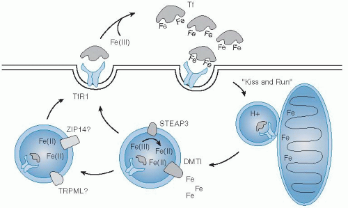
Uptake of iron by cells from the transferrin-receptor binding complex begins with its internalization by clathrin-mediated endocytosis (Fig. 10.3). Clathrin-coated vesicles deliver their cargo to acidic intracellular compartments called early endosomes. The low pH of this environment promotes release of iron and stabilizes the binding of apotransferrin
to the receptor. Together, they are recycled back to the cell surface, where apotransferrin dissociates from the receptor at neutral pH (47). Reduction of ferric iron released in the lumen of the endosome is supported by the ferrireductase Steap3 (48). Subsequent transport of ferrous iron in erythroid cells is mediated by DMT1 (49). In one model, investigators proposed that reticulocyte endosomes bearing iron-bound transferrin directly deliver cargo to mitochondria for heme biosynthesis, the “kiss-and-run hypothesis” (50).


Fig. 10.3. Transferrin (Tf) cycle. DMT1, divalent metal transporter-1; Fe, iron; STEAP3, a ferrireductase; TfR1, transferrin receptor 1; TRPML, transient receptor potential cation channel, mucolipin subfamily (a transporter); ZIP 14, a zinc transporter.

Still, other transporters may provide for iron transfer in the endosomal-lysosomal system in peripheral tissues, including Zip14 (51) and TRPML1 (52). On entering the cell cytoplasm, iron is either rapidly metabolized or stored in ferritin. A small labile iron pool exists and is in the micromolar range in most cell types. In excess, this free iron can produce reactive oxygen species that can cause cellular damage. For example, loss of iron storage as a result of tissue-specific deletion of ferritin heavy chain in mice leads to liver damage (53). Defects in transferrin’s intracellular trafficking are also known to give rise to anemia in the hbd hemoglobin-deficit mouse model as a result of mutation of Sec15l1 of the exocyst complex (54). Emerging evidence indicates that the iron-regulated myotonic dystrophy kinase-related CDC42-binding kinase α (MRCKα) may be involved in modulating transferrin-mediated iron uptake, possibly by its association with the actin cytoskeletal network and the transferrin-transferrin receptor complex (55). Clearly, the transferrin cycle must be regulated tightly and in a coordinated manner to control the distribution of iron sufficient to meet metabolic demands but limited to avoid toxicity.


Regulation of Iron Status at the Cellular Level

A major mechanism regulating iron status at the cellular level involves posttranscriptional regulation of the transferrin receptor, ferritin, and other key metabolic factors. Uptake of iron into cells is proportional to levels of the transferrin receptor and reflects the needs of the cell; otherwise, excess iron must be stored in ferritin to prevent oxidative damage, as described earlier. Transcripts for the transferrin receptor and ferritin are known to contain iron-responsive elements (IREs), stem-loop RNA structures that control stability and translation of the mRNAs, respectively (56, 57). IREs are bound by iron regulatory proteins (IRP1 and IRP2) to regulate expression of these and many other important factors in iron transport, utilization, and storage in a coordinated way (Table 10.3). Under low-iron conditions, IRPs confer translational control by binding to IREs present in the 5′ ends of iron storage or efflux proteins (ferritin and ferroportin) to reduce protein synthesis. At the same time, IRP binding to 3′ ends of mRNAs for iron uptake factors (transferrin receptor-1 and DMT1) increases message stability to enhance levels of transport into cells. Conversely, under high-iron conditions, IRP-IRE binding is lost, and synthesis for proteins involved in storage or efflux is increased, whereas iron uptake is reduced.

Only gold members can continue reading. Log In or Register to continue

Stay updated, free articles. Join our Telegram channel

Jul 27, 2016 | Posted by in PUBLIC HEALTH AND EPIDEMIOLOGY | Comments Off on Iron1

Full access? Get Clinical Tree

Get Clinical Tree app for offline access