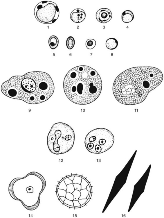
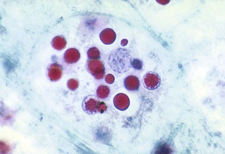
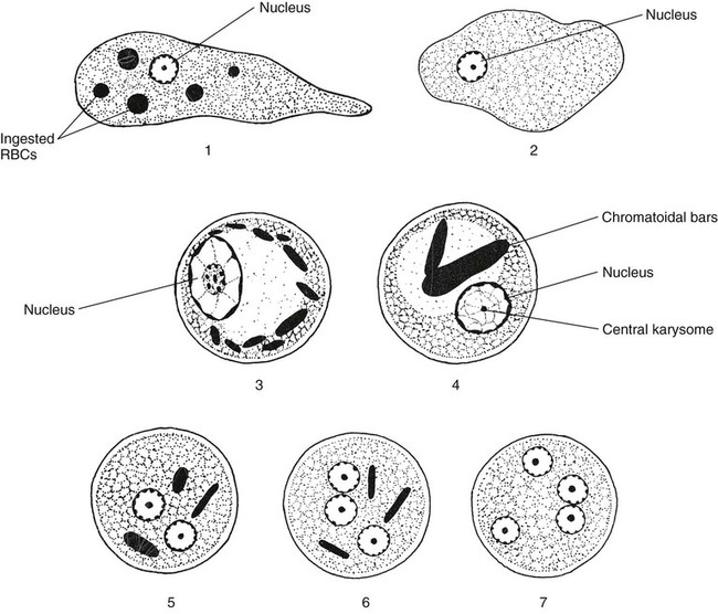
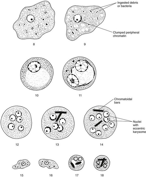
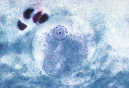
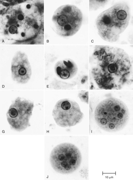
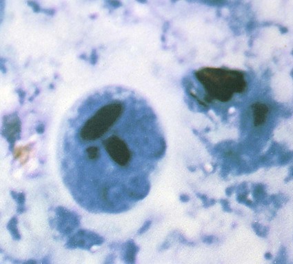
1. Describe the basic life cycle, distinguishing morphologic characteristics, clinical disease (if pathogenic), laboratory diagnosis, and prevention for the organisms listed in the following table. 2. Define and identify the following parasitic structures: trophozoite, cyst, oocyst, spore, pseudopodia, flagella, cilia, chromatoidal bars, karysome, central vacuole cyst form, axoneme, cytosotome, spiral groove, undulating membrane, ventral disc, shepherd’s crook, axostyle, macronucleus, micronucleus, apical complex, sporocyst, sporozoite, spore, sarcocyst, and polar tubule. 3. Define life cycle processes, including merogony, gametogony, sporogony, schizogony, and the associated organism or organisms and associated stages. 4. Correlate the parasitic life cycles with the specific diagnostic stages for the organisms listed. The important characteristics of the intestinal protozoa are presented in Tables 48-1 to 48-7. The clinically relevant intestinal protozoa are generally considered to be Entamoeba histolytica, Blastocystis hominis, Giardia lamblia, Dientamoeba fragilis, Balantidium coli, Isospora (Cystoisospora) belli, Cryptosporidium spp., Cyclospora cayetanensis, and the microsporidia. Nonpathogenic intestinal protozoa are listed in various figures and tables but are not discussed in detail. TABLE 48-1 Intestinal Protozoa: Trophozoites of Common Amebae *These sizes refer to wet preparation measurements. Organisms on a permanent stained smear may be 1 to 1.5 µm smaller as a result of artificial shrinkage. TABLE 48-2 Intestinal Protozoa—Cysts of Common Amebae *Wet preparation measurements; in permanent stains, organisms usually are 1 to 2 µm smaller. TABLE 48-3 Intestinal Protozoa—Trophozoites of Flagellates TABLE 48-4 Intestinal Protozoa—Cysts of Flagellates TABLE 48-5 TABLE 48-6 Morphologic Criteria Used to Identify Intestinal Protozoa (Coccidia, Blastocystis hominis) TABLE 48-7 Microsporidia That Cause Human Infection AIDS, Acquired immunodeficiency syndrome; HIV, human immunodeficiency virus; SCID, severe combined immunodeficiency *Microsporidium is a collective generic name for microsporidia that cannot be classified The class Sarcodina, or Amebae, includes the organisms capable of movement by means of cytoplasmic protrusions called pseudopodia. This group includes free-living organisms, in addition to nonpathogenic and pathogenic organisms found in the intestinal tract and other areas of the body (see Tables 48-1 and 48-2). Occasionally, when fresh stool material is examined as a direct wet mount, motile trophozoites may be seen, as well as other, nonparasitic structures (Figure 48-1). E. histolytica has directional and progressive motility, whereas the other amebae tend to move more slowly and at random. However, motility is rarely seen even in a fresh wet mount from a patient with diarrhea or dysentery. The cytoplasm is generally more finely granular, and the presence of red blood cells (RBCs) in the cytoplasm is considered diagnostic for E. histolytica (Figure 48-2). Permanent stained smears demonstrate accurate morphology compared with other techniques. When the organism is examined on a permanent stained smear (trichrome or iron-hematoxylin stain), the morphologic characteristics of E. histolytica/E. dispar are readily seen. The nucleus is characterized by evenly arranged chromatin on the nuclear membrane and a small, compact, centrally located karyosome (condensed chromatin). As mentioned, the cytoplasm usually is described as finely granular, with few ingested bacteria and scant debris in vacuoles. As stated previously, in organisms isolated from a patient with dysentery, RBCs may be visible in the cytoplasm, a feature diagnostic for E. histolytica (Figure 48-3). Most often, infection with E. histolytica is diagnosed on the basis of the organism’s morphology, without the presence of RBCs. As part of the life cycle, the trophozoites may condense into a round mass (precyst), and a thin wall is secreted around the immature cyst. Two types of inclusions may be found in this immature cyst: a glycogen mass and highly refractile chromatoidal bars (refractile chromatin structure) with smooth, rounded edges. As the cyst matures (metacyst) (see Figure 48-3; Figure 48-4), nuclear division occurs, with the production of four nuclei. Often chromatoidals may be absent in the mature cyst. Cyst morphology does not differentiate E. histolytica from E. dispar. Cyst formation occurs only in the intestinal tract; once the stool has left the body, cyst formation does not occur. The one-, two-, and four-nucleated cysts are infective and represent the mode of transmission from one host to another. Amebiasis is caused by infection with the true pathogen, Entamoeba histolytica. Recent evidence from molecular studies confirms the differentiation of pathogenic E. histolytica and nonpathogenic E. dispar (Figure 48-5) as two distinct species. E. histolytica is considered the etiologic agent of amebic colitis and extraintestinal abscesses (amebic liver abscess), whereas nonpathogenic E. dispar produces no intestinal symptoms and is not invasive in humans. E. coli trophozoites are somewhat larger than those of E. histolytica and E. dispar and range from 15 to 50 µm in diameter (see Table 48-1; Figures 48-6 and 48-7; see Figure 48-3). Motility is sluggish with broad, short pseudopods. In wet preparations, differentiating nonpathogenic E. coli from pathogenic E. histolytica is almost impossible. On the permanent stained smear viewed at a higher magnification, the cytoplasm is granular with vacuoles containing bacteria, yeasts, and other food materials. The nucleus has a large blotlike karyosome that may be eccentric rather than centrally located. The chromatin on the nuclear membrane tends to be clumped and irregular. Although rare, if RBCs are present in the intestinal tract, E. coli may ingest them rather than bacteria.
Intestinal Protozoa
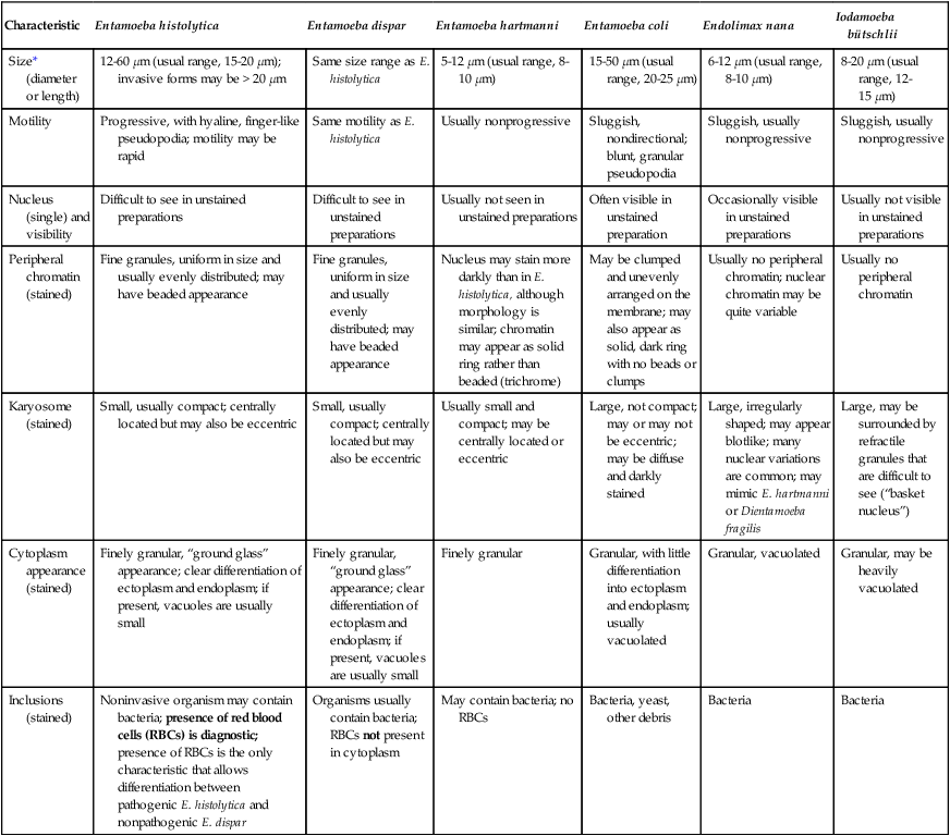
Characteristic
Entamoeba histolytica
Entamoeba dispar
Entamoeba hartmanni
Entamoeba coli
Endolimax nana
Iodamoeba bütschlii
Size* (diameter or length)
12-60 µm (usual range, 15-20 µm); invasive forms may be > 20 µm
Same size range as E. histolytica
5-12 µm (usual range, 8-10 µm)
15-50 µm (usual range, 20-25 µm)
6-12 µm (usual range, 8-10 µm)
8-20 µm (usual range, 12-15 µm)
Motility
Progressive, with hyaline, finger-like pseudopodia; motility may be rapid
Same motility as E. histolytica
Usually nonprogressive
Sluggish, nondirectional; blunt, granular pseudopodia
Sluggish, usually nonprogressive
Sluggish, usually nonprogressive
Nucleus (single) and visibility
Difficult to see in unstained preparations
Difficult to see in unstained preparations
Usually not seen in unstained preparations
Often visible in unstained preparation
Occasionally visible in unstained preparations
Usually not visible in unstained preparations
Peripheral chromatin (stained)
Fine granules, uniform in size and usually evenly distributed; may have beaded appearance
Fine granules, uniform in size and usually evenly distributed; may have beaded appearance
Nucleus may stain more darkly than in E. histolytica, although morphology is similar; chromatin may appear as solid ring rather than beaded (trichrome)
May be clumped and unevenly arranged on the membrane; may also appear as solid, dark ring with no beads or clumps
Usually no peripheral chromatin; nuclear chromatin may be quite variable
Usually no peripheral chromatin
Karyosome (stained)
Small, usually compact; centrally located but may also be eccentric
Small, usually compact; centrally located but may also be eccentric
Usually small and compact; may be centrally located or eccentric
Large, not compact; may or may not be eccentric; may be diffuse and darkly stained
Large, irregularly shaped; may appear blotlike; many nuclear variations are common; may mimic E. hartmanni or Dientamoeba fragilis
Large, may be surrounded by refractile granules that are difficult to see (“basket nucleus”)
Cytoplasm appearance (stained)
Finely granular, “ground glass” appearance; clear differentiation of ectoplasm and endoplasm; if present, vacuoles are usually small
Finely granular, “ground glass” appearance; clear differentiation of ectoplasm and endoplasm; if present, vacuoles are usually small
Finely granular
Granular, with little differentiation into ectoplasm and endoplasm; usually vacuolated
Granular, vacuolated
Granular, may be heavily vacuolated
Inclusions (stained)
Noninvasive organism may contain bacteria; presence of red blood cells (RBCs) is diagnostic; presence of RBCs is the only characteristic that allows differentiation between pathogenic E. histolytica and nonpathogenic E. dispar
Organisms usually contain bacteria; RBCs not present in cytoplasm
May contain bacteria; no RBCs
Bacteria, yeast, other debris
Bacteria
Bacteria
Characteristic
Entamoeba Histolytica/Entamoeba dispar
Entamoeba hartmanni
Entamoeba coli
Endolimax nana
Iodamoeba bütschlii
Size* (diameter or length)
10-20 µm (usual range, 12-15 µm)
5-10 µm (usual range, 6-8 µm)
10-35 µm (usual range, 15-25 µm)
5-10 µm (usual range, 6-8 µm)
5-20 µm (usual range, 10-12 µm)
Shape
Usually spherical
Usually spherical
Usually spherical; may be oval, triangular, or other shapes; may be distorted on permanent stained slide because of inadequate fixative penetration
Usually oval, may be round
May vary from oval to round; cyst may collapse because of large glycogen vacuole space
Nucleus (number and visibility)
Mature cyst: 4 nuclei
Immature cyst: 1-2 nuclei; nuclear characteristics difficult to see on wet preparation
Mature cyst: 4 nuclei; Immature cyst: 1-2 nuclei (2-nucleated cysts very common)
Mature cyst: 8 (occasionally 16 or more nuclei may be seen) Immature cysts with 2 or more nuclei are occasionally seen
Mature cyst: 4 Immature cysts: 2 Very rarely seen and may resemble cysts of Enteromonas hominis
Mature cyst: 1
Peripheral chromatin (stained)
Peripheral chromatin present; fine, uniform granules, evenly distributed; nuclear characteristics may not be as clearly visible as in trophozoite
Fine granules evenly distributed on the membrane; nuclear characteristics may be difficult to see
Coarsely granular; may be clumped and unevenly arranged on membrane; nuclear characteristics not as clearly defined as in trophozoite; may resemble E. histolytica
No peripheral chromatin
No peripheral chromatin
Karyosome (stained)
Small, compact, usually centrally located but occasionally may be eccentric
Small, compact, usually centrally located
Large, may or may not be compact and/or eccentric; occasionally may be centrally located
Smaller than karyosome seen in trophozoites but generally larger than those of genus Entamoeba
Larger, usually eccentric refractile granules may be on one side of karyosome (“basket nucleus”)
Cytoplasm, chromatoidal bodies (stained)
May be present; bodies usually elongate, with blunt, rounded, smooth edges; may be round or oval
Usually present; bodies usually elongate with blunt, rounded, smooth edges; may be round or oval
May be present (less frequently than in E. histolytica); splinter shaped with rough, pointed ends
Rare chromatoidal bodies present; occasionally small granules or inclusions seen; fine linear chromatoidals may be faintly visible on well-stained smears
No chromatoidal bodies present; occasionally small granules may be present
Glycogen (stained with iodine)
May be diffuse or absent in mature cyst; clumped chromatin mass may be present in early cysts (stains reddish brown with iodine)
May or may not be present, as in E. histolytica
May be diffuse or absent in mature cyst; clumped mass occasionally seen in mature cysts (stains reddish brown with iodine)
Usually diffuse if present (stains reddish brown with iodine)
Large, compact, well-defined mass (stains reddish brown with iodine)

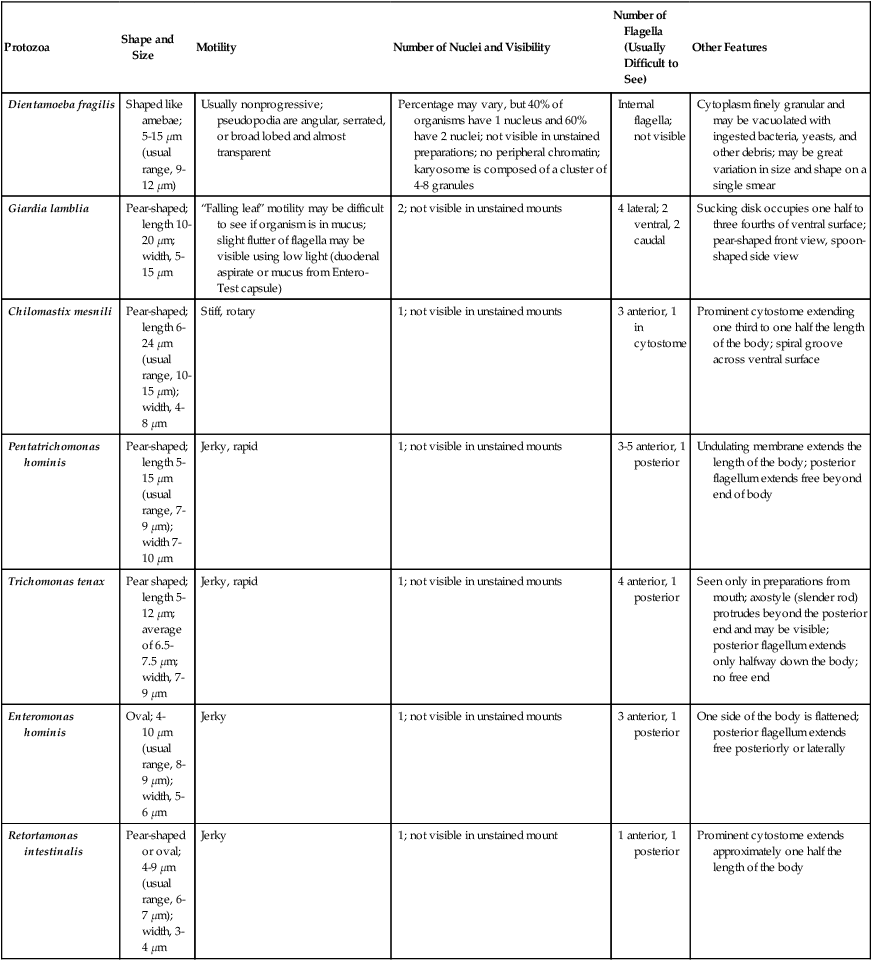
Protozoa
Shape and Size
Motility
Number of Nuclei and Visibility
Number of Flagella (Usually Difficult to See)
Other Features
Dientamoeba fragilis
Shaped like amebae; 5-15 µm (usual range, 9-12 µm)
Usually nonprogressive; pseudopodia are angular, serrated, or broad lobed and almost transparent
Percentage may vary, but 40% of organisms have 1 nucleus and 60% have 2 nuclei; not visible in unstained preparations; no peripheral chromatin; karyosome is composed of a cluster of 4-8 granules
Internal flagella; not visible
Cytoplasm finely granular and may be vacuolated with ingested bacteria, yeasts, and other debris; may be great variation in size and shape on a single smear
Giardia lamblia
Pear-shaped; length 10-20 µm; width, 5-15 µm
“Falling leaf” motility may be difficult to see if organism is in mucus; slight flutter of flagella may be visible using low light (duodenal aspirate or mucus from Entero-Test capsule)
2; not visible in unstained mounts
4 lateral; 2 ventral, 2 caudal
Sucking disk occupies one half to three fourths of ventral surface; pear-shaped front view, spoon-shaped side view
Chilomastix mesnili
Pear-shaped; length 6-24 µm (usual range, 10-15 µm); width, 4-8 µm
Stiff, rotary
1; not visible in unstained mounts
3 anterior, 1 in cytostome
Prominent cytostome extending one third to one half the length of the body; spiral groove across ventral surface
Pentatrichomonas hominis
Pear-shaped; length 5-15 µm (usual range, 7-9 µm); width 7-10 µm
Jerky, rapid
1; not visible in unstained mounts
3-5 anterior, 1 posterior
Undulating membrane extends the length of the body; posterior flagellum extends free beyond end of body
Trichomonas tenax
Pear shaped; length 5-12 µm; average of 6.5-7.5 µm; width, 7-9 µm
Jerky, rapid
1; not visible in unstained mounts
4 anterior, 1 posterior
Seen only in preparations from mouth; axostyle (slender rod) protrudes beyond the posterior end and may be visible; posterior flagellum extends only halfway down the body; no free end
Enteromonas hominis
Oval; 4-10 µm (usual range, 8-9 µm); width, 5-6 µm
Jerky
1; not visible in unstained mounts
3 anterior, 1 posterior
One side of the body is flattened; posterior flagellum extends free posteriorly or laterally
Retortamonas intestinalis
Pear-shaped or oval; 4-9 µm (usual range, 6-7 µm); width, 3-4 µm
Jerky
1; not visible in unstained mount
1 anterior, 1 posterior
Prominent cytostome extends approximately one half the length of the body
Protozoa
Size
Shape
Number of Nuclei
Other Features
Dientamoeba fragilis, Pentatrichomonas hominis, Trichomonas tenax
No cyst stage
Giardia lamblia
8-19 µm (usual range, 11-14 µm); width, 7-10 µm
Oval, ellipsoidal, or may appear round
4; not distinct in unstained preparations; usually located at one end
Longitudinal fibers in cysts may be visible in unstained preparations; deep staining median bodies usually lie across the longitudinal fibers. Shrinkage is common, with the cytoplasm pulling away from the cyst wall; “halo” effect may be seen around the outside of the cyst wall because of shrinkage caused by dehydrating reagents
Chilomastix mesnili
6-10 µm (usual range, 7-9 µm); width, 4-6 µm
Lemon or pear shaped with anterior hyaline knob
1; not distinct in unstained preparations
Cytostome with supporting fibrils, usually visible in stained preparation; curved fibril along side of cytostome, usually referred to as a “shepherd’s crook”
Enteromonas hominis
4-10 µm (usual range, 6-8 µm); width, 4-6 µm
Elongate or oval
1-4; usually 2 lying at opposite ends of cyst; not visible in unstained mounts
Resembles Endolimax nana cyst; fibrils or flagella usually not seen
Retortamonas intestinalis
4-9 µm (usual range, 4-7 µm); width, 5 µm
Pear shaped or slightly lemon shaped
1; not visible in unstained mounts
Resembles Chilomastix cyst; shadow outline of cytostome with supporting fibrils extends above nucleus; “bird beak” fibril arrangement

Protozoa
Shape and Size
Motility
Number of Nuclei
Other Features
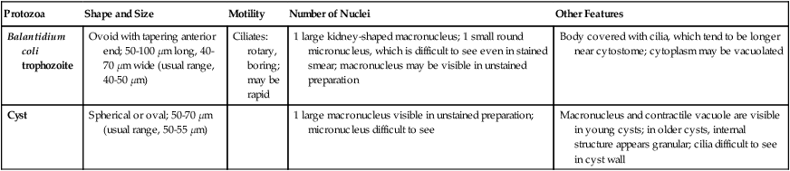
Balantidium coli
trophozoite
Ovoid with tapering anterior end; 50-100 µm long, 40-70 µm wide (usual range, 40-50 µm)
Ciliates: rotary, boring; may be rapid
1 large kidney-shaped macronucleus; 1 small round micronucleus, which is difficult to see even in stained smear; macronucleus may be visible in unstained preparation
Body covered with cilia, which tend to be longer near cytostome; cytoplasm may be vacuolated
Cyst
Spherical or oval; 50-70 µm (usual range, 50-55 µm)
1 large macronucleus visible in unstained preparation; micronucleus difficult to see
Macronucleus and contractile vacuole are visible in young cysts; in older cysts, internal structure appears granular; cilia difficult to see in cyst wall
Protozoa
Shape and Size
Other Features
Cryptosporidium spp.
C. parvum (humans and animals)
C. hominis (humans)
Oocyst generally round, 4-6 µm; each mature oocyst contains four sporozoites
Oocyst, diagnostic stage in stool, sporozoites occasionally visible within oocyst wall; acid-fast positive using modified acid-fast stains; various other stages in life cycle can be seen in biopsy specimens taken from gastrointestinal tract (brush border of epithelial cells) and other tissues; disseminated infection well documented in compromised host; oocysts immediately infective (in both formed and/or watery specimens); nosocomial infections documented; use enteric precautions for inpatients.
Cyclospora cayetanensis
Oocyst generally round, 8-10 µm; oocysts are not mature, no visible internal structure; oocysts may appear wrinkled
Oocyst, diagnostic stage in stool; acid-fast variable using modified acid-fast stains; color range from clear to deep purple (tremendous variation); best results obtained with decolorizing solution consisting of 1% acid, 3% maximum; oocysts may appear wrinkled (like crumpled cellophane); mimic Cryptosporidium oocysts but are twice as large.
Isospora (Cystoisospora) belli
Ellipsoidal oocyst; range 20-30 µm long, 10-19 µm wide; sporocysts rarely seen broken out of oocysts but measure 9-11µm
Mature oocyst contains two sporocysts with four sporozoites each; usual diagnostic stage in feces is immature oocyst containing spherical mass of protoplasm (intestinal tract). Oocysts are modified acid-fast positive. Whole oocyst may stain pink, but just the internal sporocysts stain if the oocyst is mature.
Sarcocystis hominis
S. suihominis
S. bovihominis
Oocyst thin-walled and contains two mature sporocysts, each containing four sporozoites; frequently thin oocyst wall ruptures; ovoid sporocysts each measure 10-16 µm long and 7.5-12 µm wide
Thin-walled oocyst or ovoid sporocysts occur in stool (intestinal tract)
S. “lindemanni”
Shapes and sizes of skeletal and cardiac muscle sarcocysts vary considerably
Sarcocysts contain several hundred to several thousand trophozoites, each measuring 12-16µm long and 4-9µm wide. Sarcocysts may also be divided into compartments by septa, which are not seen in Toxoplasma cysts (tissue/muscle).
Blastocystis hominis
Organisms are generally round, measure approximately 6-40 µm, and are usually characterized by a large, central body (looks like a large vacuole); this stage has been called the central body form
The more amebic form can be seen in diarrheal fluid but is difficult to identify. The central body forms vary tremendously in size, even on a single fecal smear; this is the most common form seen. Routine fecal examinations may indicate a positive rate much higher than other protozoa; some laboratories report figures of 20% and higher.
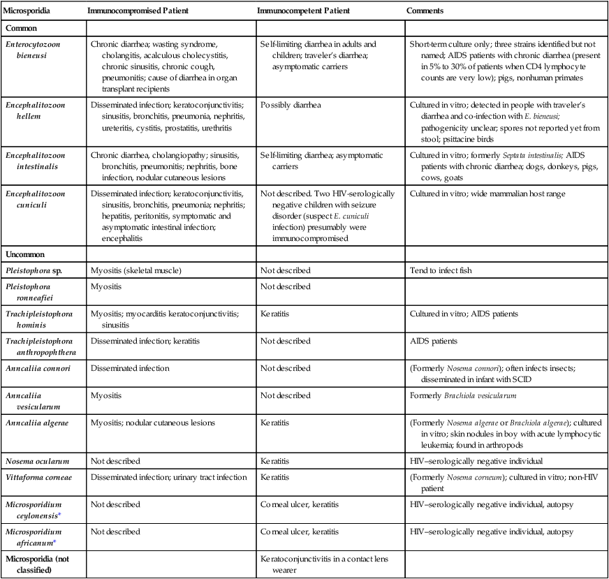
Microsporidia
Immunocompromised Patient
Immunocompetent Patient
Comments
Common
Enterocytozoon bieneusi
Chronic diarrhea; wasting syndrome, cholangitis, acalculous cholecystitis, chronic sinusitis, chronic cough, pneumonitis; cause of diarrhea in organ transplant recipients
Self-limiting diarrhea in adults and children; traveler’s diarrhea; asymptomatic carriers
Short-term culture only; three strains identified but not named; AIDS patients with chronic diarrhea (present in 5% to 30% of patients when CD4 lymphocyte counts are very low); pigs, nonhuman primates
Encephalitozoon hellem
Disseminated infection; keratoconjunctivitis; sinusitis, bronchitis, pneumonia, nephritis, ureteritis, cystitis, prostatitis, urethritis
Possibly diarrhea
Cultured in vitro; detected in people with traveler’s diarrhea and co-infection with E. bieneusi; pathogenicity unclear; spores not reported yet from stool; psittacine birds
Encephalitozoon intestinalis
Chronic diarrhea, cholangiopathy; sinusitis, bronchitis, pneumonitis; nephritis, bone infection, nodular cutaneous lesions
Self-limiting diarrhea; asymptomatic carriers
Cultured in vitro; formerly Septata intestinalis; AIDS patients with chronic diarrhea; dogs, donkeys, pigs, cows, goats
Encephalitozoon cuniculi
Disseminated infection; keratoconjunctivitis, sinusitis, bronchitis, pneumonia; nephritis; hepatitis, peritonitis, symptomatic and asymptomatic intestinal infection; encephalitis
Not described. Two HIV-serologically negative children with seizure disorder (suspect E. cuniculi infection) presumably were immunocompromised
Cultured in vitro; wide mammalian host range
Uncommon
Pleistophora sp.
Myositis (skeletal muscle)
Not described
Tend to infect fish
Pleistophora ronneafiei
Myositis
Not described
Trachipleistophora hominis
Myositis; myocarditis keratoconjunctivitis; sinusitis
Keratitis
Cultured in vitro; AIDS patients
Trachipleistophora anthropophthera
Disseminated infection; keratitis
Not described
AIDS patients
Anncaliia connori
Disseminated infection
Not described
(Formerly Nosema connori); often infects insects; disseminated in infant with SCID
Anncaliia vesicularum
Myositis
Not described
Formerly Brachiola vesicularum
Anncaliia algerae
Myositis; nodular cutaneous lesions
Keratitis
(Formerly Nosema algerae or Brachiola algerae); cultured in vitro; skin nodules in boy with acute lymphocytic leukemia; found in arthropods
Nosema ocularum
Not described
Keratitis
HIV–serologically negative individual
Vittaforma corneae
Disseminated infection; urinary tract infection
Keratitis
(Formerly Nosema corneum); cultured in vitro; non-HIV patient
Microsporidium ceylonensis*
Not described
Corneal ulcer, keratitis
HIV–serologically negative individual, autopsy
Microsporidium africanum*
Not described
Corneal ulcer, keratitis
HIV–serologically negative individual, autopsy
Microsporidia (not classified)
Keratoconjunctivitis in a contact lens wearer
Amebae
Entamoeba histolytica
General Characteristics
Epidemiology
Entamoeba coli
General Characteristics
![]()
Stay updated, free articles. Join our Telegram channel

Full access? Get Clinical Tree


Intestinal Protozoa