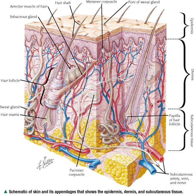
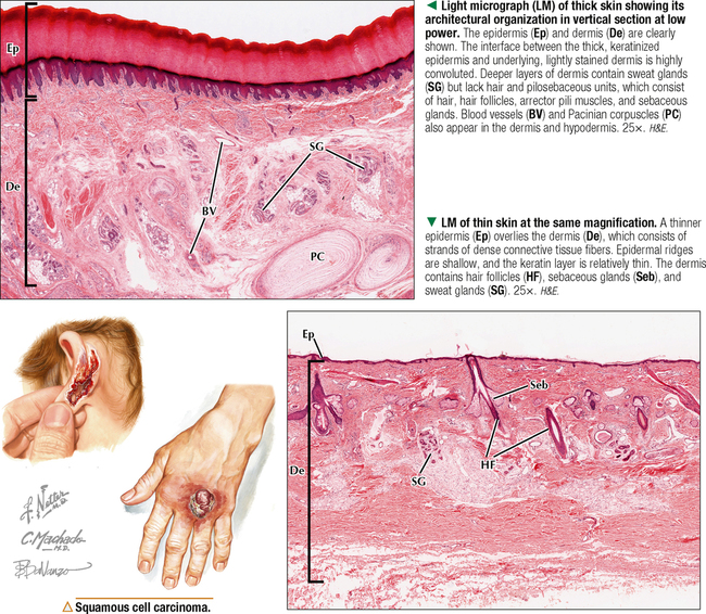
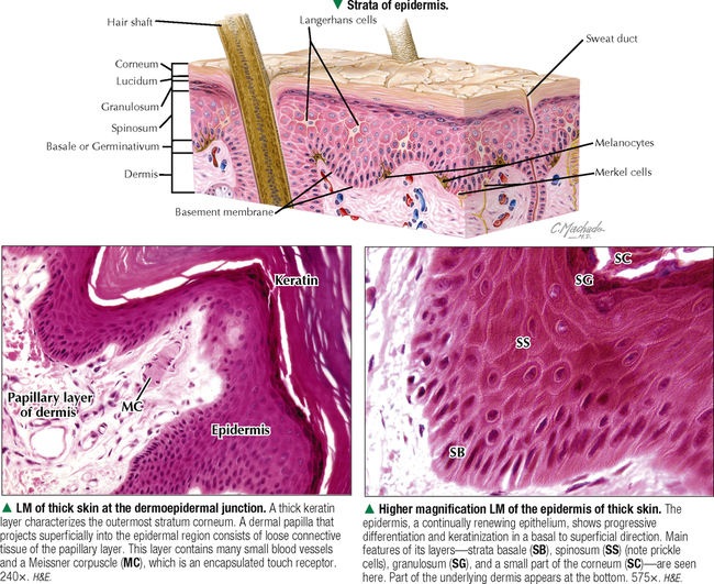
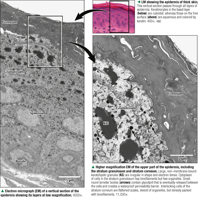
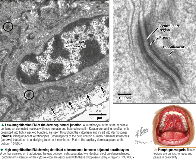
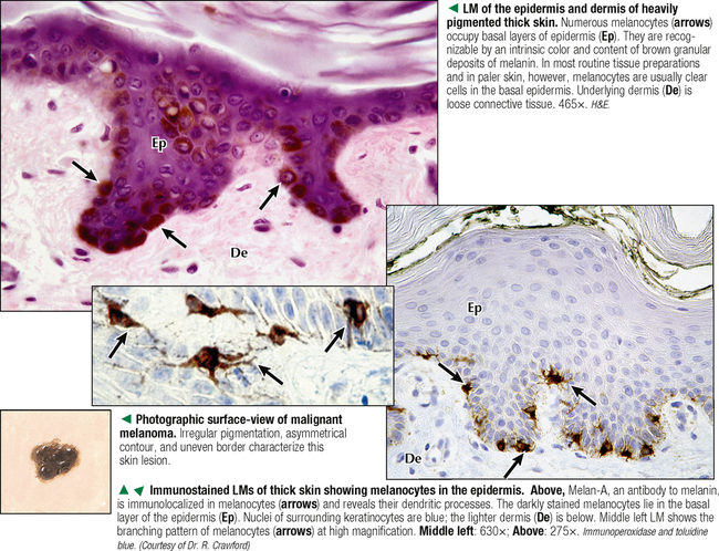
11 INTEGUMENTARY SYSTEM 11.1. Overview 11.2. Histology of Thick and Thin Skin 11.3. Histology of the Epidermis 11.4. Ultrastructure of the Epidermis 11.5. Ultrastructure of Keratinocytes 11.6. Histology and Function of Epidermal Melanocytes 11.7. Ultrastructure of Melanocytes and Melanogenesis 11.8. Structure and Function of Epidermal Langerhans Cells 11.9. Histology and Vasculature of the Dermis 11.10. Histology and Innervation of the Dermis 11.11. Histology and Function of Eccrine Sweat Glands 11.12. Histology and Function of Apocrine Sweat Glands 11.13. Histology of Pilosebaceous Units: Hair 11.14. Histology and Function of Pilosebaceous Units: Hair Follicles and Hair Growth 11.15. Ultrastructure of Hair and its Follicles 11.16. Histology of Sebaceous Glands and Arrector Pili Muscles 11.17. Ultrastructure and Function of Sebaceous Glands 11.18. Anatomy and Histology of Nails 11.19. Histology of Psoriasis 11.1 OVERVIEW The integument, the largest organ of the body, is composed of skin and skin appendages—nails, hair, sweat glands, and sebaceous glands. The total weight and overall surface area of skin in the adult are 3–5 kg and 1.5–2 m2, respectively. Skin thickness, between 0.5 and 3 mm, varies regionally; skin is thickest on the back and thinnest on the eyelid. At mucocutaneous junctions, skin is continuous with mucous membranes lining digestive, respiratory, and urogenital tracts. As well as serving as a protective barrier against injury (e.g., abrasions, cuts, burns), infectious pathogens, and ultraviolet (UV) radiation, skin assists in body temperature regulation, vitamin D synthesis, ion excretion, and sensory reception (touch and pain), and it has a remarkable regenerative capacity. It consists of stratified squamous keratinized epithelium on its outer part, called the epidermis, and an inner layer of fibrous connective tissue, called the dermis. A loose layer of subcutaneous connective tissue, the hypodermis, attaches skin to underlying structures and permits movement over most body parts. Skin has a dual embryologic origin: Epidermis and its appendages derive mostly from surface ectoderm; dermis originates from mesoderm. The epidermis consists primarily of cells called keratinocytes, which make up more than 90% of the cell population. Other epidermal cells are melanocytes and Merkel cells, which derive from neural crest, and Langerhans cells, which have a monocytic origin. During embryonic development, skin appendages deriving from the epidermis grow down into the dermis. CLINICAL POINT Cutaneous burns are classified according to depth of damage to the skin. First-degree (or superficial) burns are limited to epidermis, in which the skin presents with erythema and may peel; mild sunburn is a common example. Second-degree (or partial-thickness) burns, often caused by scalding, extend into deep (reticular) dermis, leading to inflammation, severe pain, and blister formation with little likelihood of scarring. In this case, even when most of the epithelium is destroyed, healing typically takes 1–3 weeks because of regeneration via epithelial cells surrounding hair follicles and sweat glands. More serious third-degree (or full-thickness) burns extend through the entire dermis with severe damage that may reach deeper subcutaneous layers. Because these burns are so deep, they cause little or no pain because of destruction of nerves and nerve endings. Such cases usually require special treatment (e.g., skin grafting) for healing. 11.2 HISTOLOGY OF THICK AND THIN SKIN On the basis of the structural complexity and thickness of the epidermis, skin is classified into thick or thin. Thick skin, which is glabrous, is found on palms of the hands and soles of the feet; thin skin covers most of the remaining body surface. Whereas the multilayered epidermis of thick skin is 0.8–1.5 mm thick, the epidermis of thin skin is 0.07–0.15 mm thick, with fewer cellular layers. The junction between the avascular epidermis and richly vascularized dermis—the dermoepidermal border—is usually highly corrugated and has many downward, ridge-like extensions of epidermis, called epidermal, or rete, ridges that project between alternating, upward projections of dermis, the dermal papillae. The contour of this border resembles the undersurface of an egg carton and is more complex in thick than in thin skin. A basement membrane separates epidermis from dermis. The thick dermis is divided into two layers: a superficial papillary layer of loose connective tissue containing type I and III collagen fibers interspersed with elastic fibers, connective tissue cells, and rich network of capillaries; and a deeper reticular layer of dense irregular connective tissue consisting of coarse, interlacing bundles of collagen fibers, mostly type I. Aside from fibroblasts, other connective tissue cells in the dermis include macrophages, mast cells, adipocytes, plasma cells, and lymphocytes. CLINICAL POINT Skin cancer is the most common malignant disease in North America. The three major types are basal cell carcinoma and squamous cell carcinoma (arise from keratinocytes) and melanoma (originates from melanocytes). Basal cell carcinoma accounts for more than 90% of all skin cancers; it grows slowly and seldom spreads to other parts of the body. Squamous cell carcinoma is associated with long-term exposure to sun and has a greater likelihood of metastasis. Malignant melanoma causes more than 75% of all deaths from skin cancer. If it is diagnosed early, treatment is usually effective; melanoma diagnosed at a late stage is more likely to metastasize and cause death. 11.3 HISTOLOGY OF THE EPIDERMIS The epidermis consists of cells that undergo mitosis, differentiation, maturation, and keratinization as they are displaced outward toward the skin surface to be shed. Four or five distinct layers, or strata, constitute the epidermis. The stratum basale, or germinativum, is the deepest; it consists of a single layer of closely packed, basophilic cuboidal to columnar epithelial cells, known as keratinocytes, resting on a basement membrane. These cells have oval nuclei that often show mitotic figures; they continuously undergo cell division to replace cells that move outward through the epidermis. The next layer, the stratum spinosum, is several cells thick and has polyhedral cells that become progressively flatter toward the surface. Processes of adjacent cells are attached by desmosomes. Cell shrinkage caused by a fixation artifact accentuates the processes and creates spines or prickles—thus the name prickle cells. The next layer, the stratum granulosum, consists of three to five layers of flattened cells, their axes aligned parallel to the epidermal surface. They contain numerous basophilic granules, the keratohyalin granules. Superficial to this layer is a thin, translucent, lightly eosinophilic layer, known as the stratum lucidum. Absent in thin skin but present in thick skin, it consists of a few layers of tightly packed squamous cells that lack organelles and nuclei. The outermost layer, the stratum corneum, is made of dead, anucleate cornified cells; its thickness varies regionally. The protein keratin replaces cytoplasm in its cells. The most superficial cells are continuously shed in a process known as desquamation. CLINICAL POINT Skin diseases, especially of pigmentation, are common and can result from a change in number of melanocytes or a decrease or increase in their activity. Leukoderma associated with inflammatory disorders of the skin, such as atopic dermatitis, and vitiligo are two more common hypopigmentation disorders. One of the most common hyperpigmentation disorders is melasma. It is seen primarily, but not only, in women; its onset may be during pregnancy, so it is also called mask of pregnancy. Exposure to the sun is important in induction and maintenance of hyperpigmented areas of the face. 11.4 ULTRASTRUCTURE OF THE EPIDERMIS In upper layers of the stratum spinosum, keratinocytes contain irregular, non–membrane-bound, electron-dense keratohyalin granules with diameters of 100–150 nm. These granules consist of the protein filaggrin, which cross-links with keratin. In the stratum granulosum, almost all cytoplasmic organelles and nuclei disappear because of lysosomal enzyme activity. The residual cellular profiles are filled with tightly packed filaments and are enclosed by a thickened cell membrane—the horny cell membrane. The protein involucrin binds to the inner cell membrane. Round to oval membrane-bound granules in keratinocytes in upper layers—the lamellar bodies—are 300–500 nm in diameter, are derived from Golgi complex, and are rich in glycolipids. They are eventually released from and deposited between keratinocytes, most likely forming an intercellular barrier to water. Unique keratin packing probably accounts for the presence of a stratum lucidum in plantar and palmar skin. The stratum corneum is made of interlocking cells arranged in orderly vertical stacks. These cells have thickened cell membranes and lack desmosomes, which allows cells to dissociate and desquamate easily. The normal time for turnover of keratinocytes from stratum basale to uppermost stratum corneum varies from 20 to 75 days. Turnover and transit times may be even more rapid in some diseases, such as psoriasis, in which transit time is about 8 days. 11.5 ULTRASTRUCTURE OF KERATINOCYTES Cells of the stratum basale have relatively euchromatic nuclei compared with those of more superficial layers. Their cytoplasm contains many ribosomes, mitochondria, and an extensive cytoskeleton of 10-nm intermediate filaments known as tonofilaments. These are made of the keratin family of intermediate filament proteins. All epithelial cells contain keratins, and almost 50 different types of keratins are found in skin. Keratinocytes of the strata basale and spinosum are connected by desmosomes. These complex intercellular junctions mediate and enhance cell adhesion by anchoring keratin filaments to keratinocyte plasma membranes. By linking tonofilament bundles of adjacent cells, desmosomes provide the epidermis with structural continuity and mechanical strength. To further counteract mechanical forces, basal aspects of keratinocytes are firmly attached to underlying basement membrane by hemidesmosomes. Hemidesmosomes have only one intracytoplasmic attachment plaque to which tonofilaments from the cell interior attach. Fine anchoring filaments radiate from the outer aspect of the plasma membrane into the basal lamina. The basement membrane at the dermoepidermal junction usually requires special light microscopic techniques to be visible. This specialized supporting zone of extracellular matrix consists of several layers. A lamina lucida and lamina densa together constitute the basal lamina, which contains type IV collagen, laminin, fibronectin, and proteoglycans. A deeper reticular lamina, made mainly of type I collagen fibers, merges with underlying connective tissue. CLINICAL POINT Some debilitating blistering disorders of skin result from disrupted epidermal adhesion and attachment. Antigens for these diseases are components of either desmosomes or hemidesmosomes and belong to three genetic families—cadherin, armadillo, and plakin. Autoantibodies may react with the keratinocyte cell surface or epidermal basement membrane, which induces separation of epidermal keratinocytes or dermoepidermal junctions. Pemphigus is the most common disease with anti-keratinocyte cell surface antibodies; the related bullous pemphigoid causes subepidermal blisters. In these diseases, mutations in genes encoding desmosomal components have been identified, which may lead to novel, efficient treatment strategies. 11.6 HISTOLOGY AND FUNCTION OF EPIDERMAL MELANOCYTES Melanocytes are melanin pigment-producing cells that determine color of skin and hair. The major determinant of color is not melanocyte number but activity, which is affected by corticotropin from the pituitary. Derived from the neural crest, melanocytes migrate to the basal layer of the epidermis Only gold members can continue reading. Log In or Register to continue Share this: Share on X (Opens in new window) X Share on Facebook (Opens in new window) Facebook Like this:Like Loading… Related Related posts: CARDIOVASCULAR SYSTEM SPECIAL SENSES THE CELL FEMALE REPRODUCTIVE SYSTEM Stay updated, free articles. Join our Telegram channel Join Tags: Netters Essential Histology Jun 18, 2016 | Posted by admin in HISTOLOGY | Comments Off on INTEGUMENTARY SYSTEM Full access? Get Clinical Tree
11 INTEGUMENTARY SYSTEM 11.1. Overview 11.2. Histology of Thick and Thin Skin 11.3. Histology of the Epidermis 11.4. Ultrastructure of the Epidermis 11.5. Ultrastructure of Keratinocytes 11.6. Histology and Function of Epidermal Melanocytes 11.7. Ultrastructure of Melanocytes and Melanogenesis 11.8. Structure and Function of Epidermal Langerhans Cells 11.9. Histology and Vasculature of the Dermis 11.10. Histology and Innervation of the Dermis 11.11. Histology and Function of Eccrine Sweat Glands 11.12. Histology and Function of Apocrine Sweat Glands 11.13. Histology of Pilosebaceous Units: Hair 11.14. Histology and Function of Pilosebaceous Units: Hair Follicles and Hair Growth 11.15. Ultrastructure of Hair and its Follicles 11.16. Histology of Sebaceous Glands and Arrector Pili Muscles 11.17. Ultrastructure and Function of Sebaceous Glands 11.18. Anatomy and Histology of Nails 11.19. Histology of Psoriasis 11.1 OVERVIEW The integument, the largest organ of the body, is composed of skin and skin appendages—nails, hair, sweat glands, and sebaceous glands. The total weight and overall surface area of skin in the adult are 3–5 kg and 1.5–2 m2, respectively. Skin thickness, between 0.5 and 3 mm, varies regionally; skin is thickest on the back and thinnest on the eyelid. At mucocutaneous junctions, skin is continuous with mucous membranes lining digestive, respiratory, and urogenital tracts. As well as serving as a protective barrier against injury (e.g., abrasions, cuts, burns), infectious pathogens, and ultraviolet (UV) radiation, skin assists in body temperature regulation, vitamin D synthesis, ion excretion, and sensory reception (touch and pain), and it has a remarkable regenerative capacity. It consists of stratified squamous keratinized epithelium on its outer part, called the epidermis, and an inner layer of fibrous connective tissue, called the dermis. A loose layer of subcutaneous connective tissue, the hypodermis, attaches skin to underlying structures and permits movement over most body parts. Skin has a dual embryologic origin: Epidermis and its appendages derive mostly from surface ectoderm; dermis originates from mesoderm. The epidermis consists primarily of cells called keratinocytes, which make up more than 90% of the cell population. Other epidermal cells are melanocytes and Merkel cells, which derive from neural crest, and Langerhans cells, which have a monocytic origin. During embryonic development, skin appendages deriving from the epidermis grow down into the dermis. CLINICAL POINT Cutaneous burns are classified according to depth of damage to the skin. First-degree (or superficial) burns are limited to epidermis, in which the skin presents with erythema and may peel; mild sunburn is a common example. Second-degree (or partial-thickness) burns, often caused by scalding, extend into deep (reticular) dermis, leading to inflammation, severe pain, and blister formation with little likelihood of scarring. In this case, even when most of the epithelium is destroyed, healing typically takes 1–3 weeks because of regeneration via epithelial cells surrounding hair follicles and sweat glands. More serious third-degree (or full-thickness) burns extend through the entire dermis with severe damage that may reach deeper subcutaneous layers. Because these burns are so deep, they cause little or no pain because of destruction of nerves and nerve endings. Such cases usually require special treatment (e.g., skin grafting) for healing. 11.2 HISTOLOGY OF THICK AND THIN SKIN On the basis of the structural complexity and thickness of the epidermis, skin is classified into thick or thin. Thick skin, which is glabrous, is found on palms of the hands and soles of the feet; thin skin covers most of the remaining body surface. Whereas the multilayered epidermis of thick skin is 0.8–1.5 mm thick, the epidermis of thin skin is 0.07–0.15 mm thick, with fewer cellular layers. The junction between the avascular epidermis and richly vascularized dermis—the dermoepidermal border—is usually highly corrugated and has many downward, ridge-like extensions of epidermis, called epidermal, or rete, ridges that project between alternating, upward projections of dermis, the dermal papillae. The contour of this border resembles the undersurface of an egg carton and is more complex in thick than in thin skin. A basement membrane separates epidermis from dermis. The thick dermis is divided into two layers: a superficial papillary layer of loose connective tissue containing type I and III collagen fibers interspersed with elastic fibers, connective tissue cells, and rich network of capillaries; and a deeper reticular layer of dense irregular connective tissue consisting of coarse, interlacing bundles of collagen fibers, mostly type I. Aside from fibroblasts, other connective tissue cells in the dermis include macrophages, mast cells, adipocytes, plasma cells, and lymphocytes. CLINICAL POINT Skin cancer is the most common malignant disease in North America. The three major types are basal cell carcinoma and squamous cell carcinoma (arise from keratinocytes) and melanoma (originates from melanocytes). Basal cell carcinoma accounts for more than 90% of all skin cancers; it grows slowly and seldom spreads to other parts of the body. Squamous cell carcinoma is associated with long-term exposure to sun and has a greater likelihood of metastasis. Malignant melanoma causes more than 75% of all deaths from skin cancer. If it is diagnosed early, treatment is usually effective; melanoma diagnosed at a late stage is more likely to metastasize and cause death. 11.3 HISTOLOGY OF THE EPIDERMIS The epidermis consists of cells that undergo mitosis, differentiation, maturation, and keratinization as they are displaced outward toward the skin surface to be shed. Four or five distinct layers, or strata, constitute the epidermis. The stratum basale, or germinativum, is the deepest; it consists of a single layer of closely packed, basophilic cuboidal to columnar epithelial cells, known as keratinocytes, resting on a basement membrane. These cells have oval nuclei that often show mitotic figures; they continuously undergo cell division to replace cells that move outward through the epidermis. The next layer, the stratum spinosum, is several cells thick and has polyhedral cells that become progressively flatter toward the surface. Processes of adjacent cells are attached by desmosomes. Cell shrinkage caused by a fixation artifact accentuates the processes and creates spines or prickles—thus the name prickle cells. The next layer, the stratum granulosum, consists of three to five layers of flattened cells, their axes aligned parallel to the epidermal surface. They contain numerous basophilic granules, the keratohyalin granules. Superficial to this layer is a thin, translucent, lightly eosinophilic layer, known as the stratum lucidum. Absent in thin skin but present in thick skin, it consists of a few layers of tightly packed squamous cells that lack organelles and nuclei. The outermost layer, the stratum corneum, is made of dead, anucleate cornified cells; its thickness varies regionally. The protein keratin replaces cytoplasm in its cells. The most superficial cells are continuously shed in a process known as desquamation. CLINICAL POINT Skin diseases, especially of pigmentation, are common and can result from a change in number of melanocytes or a decrease or increase in their activity. Leukoderma associated with inflammatory disorders of the skin, such as atopic dermatitis, and vitiligo are two more common hypopigmentation disorders. One of the most common hyperpigmentation disorders is melasma. It is seen primarily, but not only, in women; its onset may be during pregnancy, so it is also called mask of pregnancy. Exposure to the sun is important in induction and maintenance of hyperpigmented areas of the face. 11.4 ULTRASTRUCTURE OF THE EPIDERMIS In upper layers of the stratum spinosum, keratinocytes contain irregular, non–membrane-bound, electron-dense keratohyalin granules with diameters of 100–150 nm. These granules consist of the protein filaggrin, which cross-links with keratin. In the stratum granulosum, almost all cytoplasmic organelles and nuclei disappear because of lysosomal enzyme activity. The residual cellular profiles are filled with tightly packed filaments and are enclosed by a thickened cell membrane—the horny cell membrane. The protein involucrin binds to the inner cell membrane. Round to oval membrane-bound granules in keratinocytes in upper layers—the lamellar bodies—are 300–500 nm in diameter, are derived from Golgi complex, and are rich in glycolipids. They are eventually released from and deposited between keratinocytes, most likely forming an intercellular barrier to water. Unique keratin packing probably accounts for the presence of a stratum lucidum in plantar and palmar skin. The stratum corneum is made of interlocking cells arranged in orderly vertical stacks. These cells have thickened cell membranes and lack desmosomes, which allows cells to dissociate and desquamate easily. The normal time for turnover of keratinocytes from stratum basale to uppermost stratum corneum varies from 20 to 75 days. Turnover and transit times may be even more rapid in some diseases, such as psoriasis, in which transit time is about 8 days. 11.5 ULTRASTRUCTURE OF KERATINOCYTES Cells of the stratum basale have relatively euchromatic nuclei compared with those of more superficial layers. Their cytoplasm contains many ribosomes, mitochondria, and an extensive cytoskeleton of 10-nm intermediate filaments known as tonofilaments. These are made of the keratin family of intermediate filament proteins. All epithelial cells contain keratins, and almost 50 different types of keratins are found in skin. Keratinocytes of the strata basale and spinosum are connected by desmosomes. These complex intercellular junctions mediate and enhance cell adhesion by anchoring keratin filaments to keratinocyte plasma membranes. By linking tonofilament bundles of adjacent cells, desmosomes provide the epidermis with structural continuity and mechanical strength. To further counteract mechanical forces, basal aspects of keratinocytes are firmly attached to underlying basement membrane by hemidesmosomes. Hemidesmosomes have only one intracytoplasmic attachment plaque to which tonofilaments from the cell interior attach. Fine anchoring filaments radiate from the outer aspect of the plasma membrane into the basal lamina. The basement membrane at the dermoepidermal junction usually requires special light microscopic techniques to be visible. This specialized supporting zone of extracellular matrix consists of several layers. A lamina lucida and lamina densa together constitute the basal lamina, which contains type IV collagen, laminin, fibronectin, and proteoglycans. A deeper reticular lamina, made mainly of type I collagen fibers, merges with underlying connective tissue. CLINICAL POINT Some debilitating blistering disorders of skin result from disrupted epidermal adhesion and attachment. Antigens for these diseases are components of either desmosomes or hemidesmosomes and belong to three genetic families—cadherin, armadillo, and plakin. Autoantibodies may react with the keratinocyte cell surface or epidermal basement membrane, which induces separation of epidermal keratinocytes or dermoepidermal junctions. Pemphigus is the most common disease with anti-keratinocyte cell surface antibodies; the related bullous pemphigoid causes subepidermal blisters. In these diseases, mutations in genes encoding desmosomal components have been identified, which may lead to novel, efficient treatment strategies. 11.6 HISTOLOGY AND FUNCTION OF EPIDERMAL MELANOCYTES Melanocytes are melanin pigment-producing cells that determine color of skin and hair. The major determinant of color is not melanocyte number but activity, which is affected by corticotropin from the pituitary. Derived from the neural crest, melanocytes migrate to the basal layer of the epidermis Only gold members can continue reading. Log In or Register to continue Share this: Share on X (Opens in new window) X Share on Facebook (Opens in new window) Facebook Like this:Like Loading… Related Related posts: CARDIOVASCULAR SYSTEM SPECIAL SENSES THE CELL FEMALE REPRODUCTIVE SYSTEM Stay updated, free articles. Join our Telegram channel Join Tags: Netters Essential Histology Jun 18, 2016 | Posted by admin in HISTOLOGY | Comments Off on INTEGUMENTARY SYSTEM Full access? Get Clinical Tree