Immune System


Immune System


The immune system is responsible for safe-guarding the body from disease-causing microorganisms. It’s part of a complex system of host defenses.

Host defenses may be innate or acquired. Innate defenses include physical and chemical barriers, the complement complex, and cells, such as phagocytes (cells programmed to destroy foreign cells, such as bacteria) and natural killer lymphocytes.

Physical barriers, such as the skin and mucous membranes, prevent invasion by most organisms. Chemical barriers include lysozymes (found in such body secretions as tears, mucus, and saliva) and hydrochloric acid in the stomach. Lysozymes destroy bacteria by removing cell walls. Hydrochloric acid breaks down foods and destroys pathogens carried by food or swallowed mucus.

Organisms that penetrate this first line of defense simultaneously trigger the inflammatory and immune responses, some innate and others acquired.

Acquired immunity comes into play when the body encounters a cell or cell product that it recognizes as foreign, such as a bacterium or a virus. The two types of immunity provided by cells are humoral (provided by B lymphocytes) and cell mediated (provided by T lymphocytes). All cells involved in the inflammatory and immune responses arrive from a single type of stem cell in the bone marrow. B cells mature in the marrow, and T cells migrate to the thymus, where they mature.

The inflammatory response is the immediate local response to tissue injury, whether from trauma or infection. It involves the action of polymorphonuclear leukocytes, basophils and mast cells, platelets and, to some extent, monocytes and macrophages. Each of these cells is described in a later section of this chapter.


Immune response

The immune response primarily involves the interaction of antigens (foreign proteins), B lymphocytes, T lymphocytes, macrophages, cytokines, complement, and polymorphonuclear leukocytes. Some immunoactive cells circulate constantly; others remain in the tissues and organs of the immune system, such as the thymus, lymph nodes, bone marrow, spleen, and tonsils. In the thymus, the T lymphocytes, which are involved in cell-mediated immunity, become able to differentiate self (host) from nonself (foreign) substances (antigens). In contrast, B lymphocytes, which are involved in humoral immunity, mature in the bone marrow. The key mechanism in humoral immunity is the production of immunoglobulin by B cells and the subsequent activation of the complement cascade. The lymph nodes, spleen, liver, and intestinal lymphoid tissue help remove and destroy circulating antigens in the blood and lymph.

image The immune system’s ability to fight off infections and other immune system disorders decreases with age.

Fewer lymphocytes are present, and the ones that are there are less responsive to invasion of the body by infection and other antigens.


Autoantibodies, causative factors in such diseases as rheumatoid arthritis and atherosclerosis, are more likely to occur with aging.

Organs, such as the thymus gland, are less efficient in producing hormones related to the immune system.

image At age 2, a child’s immune system is fully functioning. Infants between ages 6 and 9 months are particularly vulnerable to disease because they’re no longer supported by maternal antibodies and their own immune system isn’t yet established.


ANTIGENS

An antigen is a substance that can induce an immune response. T and B lymphocytes have specific receptors that respond to specific antigen molecular shapes, called epitopes. In B cells, this receptor is an immunoglobulin, also called an antibody.


Major histocompatibility complex

The T-cell antigen receptor recognizes antigens only in association with specific cell-surface molecules known as the major histocompatibility complex (MHC).

The MHC, also known as the human leukocyte antigen (HLA) locus, is a cluster of genes on human chromosome 6 that has a pivotal role in the immune response. Every person receives one set of MHC genes from each parent, and both sets of genes are expressed on the individual’s cells. These genes produce MHC molecules, which participate in:

♦ the recognition of self versus nonself

♦ the interaction of immunologically active cells by coding for cell-surface proteins.

MHC molecules differ among individuals. Slightly different antigen receptors can recognize a large number of distinct antigens, coded by distinct, variable region genes.

Groups or clones of lymphocytes that have identical receptors for a specific antigen exist. The clone of a lymphocyte rapidly proliferates when exposed to the specific antigen. Some lymphocytes further differentiate, whereas others become memory cells, which allow a more rapid response—the memory, or anamnestic, response—to subsequent challenge by the antigen.


Haptens

Most antigens are large molecules, such as proteins or polysaccharides. Smaller molecules, such as drugs, that aren’t antigenic by themselves are known as haptens. They can bind with larger molecules, or carriers, and become antigenic or immunogenic.


Antigenicity

Many factors influence the intensity of a foreign substance’s interaction with the host’s immune system (antigenicity):

♦ physical and chemical characteristics of the antigen

♦ its relative foreignness—for example, little or no immune response may follow the transfusion of serum proteins between humans, but a vigorous immune response (serum sickness) commonly follows transfusion of horse serum proteins to a human

♦ the host’s genetic makeup, especially the MHC molecules.


HUMORAL IMMUNITY

The humoral immune response is one of two types of immune responses that can occur when foreign substances invade the body. The other is the cell-mediated response. The humoral response is also called an antibodymediated response.


B lymphocytes

B lymphocytes and their products, immunoglobulins, are the basis of humoral immunity. A soluble antigen binds with the B-cell antigen receptor, initiating the humoral immune response. The activated B cells differentiate into plasma cells, which secrete immunoglobulins, also called antibodies. This response is regulated by T lymphocytes and their products—lymphokines, such as interleukin-2 (IL-2), IL-4, IL-5, and interferon-8—which determine which class of immunoglobulins a B cell will manufacture.


Immunoglobulins

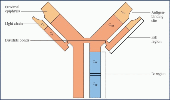
The immunoglobulins that plasma cells secrete are four-chain molecules with two heavy and two light chains. Each chain has a variable (V) region and one or more constant (C) regions, which are coded by separate genes. The V regions of both light and heavy chains participate in antigen binding. The C regions of the heavy chain provide a binding site for crystallizable fragment (Fc) receptors on cells and govern other mechanisms. (See Structure of the immunoglobulin molecule.)

There are five known classes of immunoglobulins: IgG, IgM, IgA, IgE, and IgD. These are distinguished by the constant portions of their heavy chains. However, each class has a kappa or lambda light chain, which gives rise to many subtypes and provides almost limitless combinations of light and heavy chains that give immunoglobulins their specificity. (See Classification of immunoglobulins, page 436.)


A clone of B cells is specific for only one antigen, and the V regions of its Ig light chains determines that specificity. However, the class of immunoglobulin can change if the association between the cell’s V region genes and heavy chain C region genes changes through a process known as isotype switching. For example, a clone of B cells genetically programmed to recognize tetanus toxoid will first make an IgM antibody against tetanus toxoid and later an IgG or other antibody against it.


CELL-MEDIATED IMMUNITY

The cell-mediated immune response protects the body against bacterial, viral, and fungal infections and defends against transplanted cells and tumor cells. T lymphocytes and macrophages are the chief participants in the cell-mediated immune response. A macrophage processes the antigen and then presents it to T lymphocytes.


Macrophages

Macrophages influence both immune and inflammatory responses. Macrophage precursors circulate in the blood. When they collect in various tissues and organs, they differentiate into different types of macrophages. Unlike B and T lymphocytes, macrophages lack surface receptors for specific antigens. Instead, they have receptors for the C region of the heavy chain (Fc region) of immunoglobulin, for fragments of the third component of complement (C3), and for nonimmunologic substances, such as carbohydrate molecules.

One of the most important functions of macrophages is presentation of antigen to T lymphocytes. Macrophages ingest and process the antigen, then they deposit it on their own surfaces in association with HLA antigen. T lymphocytes become activated when they recognize the antigen-HLA complex. Macrophages also function in the inflammatory response by producing IL-1, which generates fever, and by
synthesizing complement proteins and other mediators that have phagocytic, microbicidal, and oncolytic effects.


T lymphocytes

Immature T lymphocytes are derived from the bone marrow and migrate to the thymus, where they mature. In maturation, the products of the MCH genes “teach” T cells to distinguish between self and nonself.

Five types of T cells exist with specific functions:

♦ memory cells, sensitized cells that remain dormant until second exposure to antigen, also known as secondary immune response

♦ lymphokine-producing cells, delayed hypersensitivity reactions

♦ cytotoxic T cells, direct destruction of antigen or the cells carrying the antigen

♦ helper T cells, also known as T4 cells, facilitate the humoral and cell-mediated responses

♦ suppressor T cells, also known as T8 cells, inhibit humoral and cell-mediated responses.

T cells acquire specific surface molecules (markers) that identify their potential role when needed in the immune response. These markers and the T-cell antigen receptor together promote the particular activation of each type of T cell. T-cell activation requires presentation of antigens in the context of a specific HLA antigen—for example, class II HLA for helper T cells and class I HLA for cytotoxic T cells. T-cell activation also requires IL-1, produced by macrophages, and IL-2, produced by T cells.


Natural killer cells

Natural killer cells make up a discrete population of large lymphocytes, some of which resemble
T cells. They recognize surface changes on body cells infected with a virus. They bind to and, in many cases, kill the infected cells.


Cytokines

Cytokines are low-molecular-weight proteins involved in the communication among macrophages and the lymphocytes. They induce or regulate various immune or inflammatory responses. Cytokines include colony-stimulating factors, interferons, interleukins, tumor necrosis factors, and transforming growth factor.


COMPLEMENT SYSTEM

The complement system, the chief humoral effector of the inflammatory response, includes more than 20 serum proteins. When activated, these proteins interact in a cascade-like process that has profound biological effects. Complement activation takes place through one of two pathways.


Classic pathway

In the classic pathway, IgM or IgG binds with the antigen to form antigen-antibody complexes that activate the first complement component, C1. This in turn activates C4, C2, and C3.


Alternate pathway

In the alternate pathway, activating surfaces, such as bacterial cell membranes, directly amplify spontaneous cleavage of C3. Once C3 is activated in either pathway, activation of the terminal components, C5 to C9, follows.

The major biological effects of complement activation include chemotaxis (phagocyte attraction), phagocyte activation, histamine release, viral neutralization, promotion of phagocytosis by opsonization (making the bacteria susceptible to phagocytosis), and lysis of cells and bacteria. Kinins (peptides that cause vasodilation and enhance vascular permeability and smooth-muscle contraction) and other mediators of inflammation derived from the kinin and coagulation pathways interact with the complement system.


POLYMORPHONUCLEAR LEUKOCYTES

Other key factors in the inflammatory response are the polymorphonuclear leukocytes: neutrophils, eosinophils, basophils, and mast cells.


Neutrophils

Neutrophils, the most numerous of these leukocytes, derive from bone marrow and increase dramatically in number in response to infection and inflammation. They’re the first to respond in acute infection. Neutrophils are highly mobile cells attracted to areas of inflammation and are the main constituent of pus.

Neutrophils have surface receptors for immunoglobulins and complement fragments, and they avidly ingest bacteria or other particles that are coated with target-identifying antibodies (opsonins). Toxic oxygen metabolites and enzymes such as lysozyme promptly kill the ingested organisms. Unfortunately, in addition to killing invading organisms, neutrophils also damage host tissues.


Eosinophils

Eosinophils, also derived from bone marrow, multiply in allergic and parasitic disorders. Although their phagocytic function isn’t clearly understood, evidence suggests that they participate in host defense against parasites. Their products may also diminish inflammatory response in allergic disorders.


Basophils and mast cells

Basophils and mast cells also function in immune disorders. Mast cells, unlike basophils, aren’t blood cells. Basophils circulate in peripheral blood, whereas mast cells accumulate in connective tissue, particularly in the lungs, intestines, and skin. Both types of cells have surface receptors for IgE. When their receptors are cross-linked by an IgE antigen complex, they release mediators characteristic of the allergic response.


Pathophysiologic changes

The host defense system and the immune response are highly complex processes, subject to malfunction at any point along the sequence of events. This malfunction may involve exaggeration, misdirection, or an absence or depression of activity leading to an immune disorder.


IMMUNE RESPONSE MALFUNCTION

When the immune system responds inappropriately, three basic categories of reactions may occur: hypersensitivity, autoimmune response, and alloimmune response. The type of reaction is determined by the source of the antigen, such as environmental, self, or other person, to which the immune system is responding.


Hypersensitivity

Hypersensitivity is an exaggerated or inappropriate response that occurs on second exposure to an antigen. The result is inflammation and the destruction of healthy tissue. Allergy refers
to the harmful effects resulting from a hypersensitivity to antigens, also called allergens.

Hypersensitivity reactions may be immediate, occurring within minutes to hours of reexposure, or delayed, occurring several hours after reexposure. A delayed hypersensitivity reaction typically is most severe days after the reexposure.

Generally, hypersensitivity reactions are classified as one of four types: type I (IgE mediated), type II (tissue specific), type III (immune complex mediated), type IV (cell mediated).


Type I hypersensitivity (allergic disorders)

With type I hypersensitivity, allergens activate T cells, which induce B-cell production of IgE, which binds to the Fc receptors on the surface of mast cells. Repeated exposure to relatively large doses of the allergen is usually necessary to cause this response. When enough IgE has been produced, the person is sensitized to the allergen. At the next exposure to the same antigen, the antigen binds with the surface IgE, cross-links the Fc receptors, and causes mast cells to degranulate and release various mediators. Degranulation also may be triggered by complement-driven anaphylatoxins—C3a and C5a—or by certain drugs, such as morphine.

Some of the mediators released are preformed, whereas others are newly synthesized on activation of the mast cells. Preformed mediators include heparin, histamine, proteolytic (protein-splitting) and other enzymes, and chemotactic factors for eosinophils and neutrophils. Newly synthesized mediators include prostaglandins and leukotrienes. Mast cells also produce various cytokines, which initiate smooth-muscle contraction, vasodilation, bronchospasm, edema, increased vascular permeability, mucus secretion, and cellular infiltration by eosinophils and neutrophils. These effects result in some of the classic associated signs and symptoms, such as hypotension, wheezing, swelling, urticaria, and rhinorrhea.


Type II hypersensitivity (antibody-dependent cytotoxicity)

A tissue-specific reaction, type II hypersensitivity, generally involves the destruction of a target cell by an antibody directed against cell-surface antigens. Alternatively, the antibody may be directed against small molecules adsorbed to cells or against cell-surface receptors, rather than against the cell constituents themselves. Tissue damage occurs through several mechanisms:

♦ binding of antigen and antibody activates complement, which ultimately disrupts cellular membranes—complement-mediated lysis

♦ various phagocytic cells with receptors for immunoglobulin (Fc region) and complement fragments envelop and destroy opsonized targets, such as red blood cells, leukocytes, and platelets

♦ cytotoxic T cells and natural killer cells, though not antigen specific, also contribute to tissue damage by releasing toxic substances that destroy the cells

♦ antibody binding causes the target cell to malfunction rather than causing its destruction.


Type III hypersensitivity (immune complex disease)

In type III hypersensitivity, circulating antigenantibody complexes (immune complexes) accumulate and are deposited in the tissues. The most common tissues involved are the kidneys, joints, skin, and blood vessels. Normally, they clear excess immune complexes from the circulation. However, immune complexes deposited in the tissues activate the complement cascade, causing local inflammation, and trigger platelet release of vasoactive amines that increase vascular permeability, so more immune complexes accumulate in the vessel walls.

Probably the most harmful effects result from the generation of complement fragments that attract neutrophils. The neutrophils attempt to ingest the immune complexes. They’re generally unsuccessful, but in the attempt, the neutrophils release lysosomal enzymes, which exacerbate the tissue damage.

The formation of immune complexes is dynamic and ever changing. The complexes that form in children may be totally different from those formed in adolescents and adults. Also, more than one type of immune complex may be present at one time.


Type IV hypersensitivity (delayed hypersensitivity)

Type IV hypersensitivity cell-mediated reactions involve the processing of the antigen by the macrophages. Once processed, the antigen is presented to the T cells. Cytotoxic T cells, if activated, attack and destroy the target cells directly. When lymphokine T cells are activated, they release lymphokines, which recruit and activate other lymphocytes, monocytes, macrophages, and polymorphonuclear leukocytes. The coagulation, kinin, and complement cascades also contribute to tissue damage in this type of reaction.


Autoimmune reactions

In autoimmune reactions, the body’s normal defenses become self-destructive, recognizing
self-antigens as foreign. What causes this misdirected response isn’t clearly understood. For example, drugs or viruses have been implicated as causing some autoimmune reactions, but in such diseases as rheumatoid arthritis and systemic lupus erythematosus the mechanism for misdirection is unclear.

Autoimmune reactions are believed to result from a combination of factors, including genetic, hormonal, and environmental influences. Many are characterized by B-cell hyperactivity and by hypergammaglobulinemia. B-cell hyperactivity may be related to T-cell abnormalities. Hormonal and genetic factors strongly influence the onset of some autoimmune disorders.


Alloimmune reactions

Alloimmune reactions are directed at antigens from the tissues of others of the same species. These reactions commonly occur in transplant and transfusion reactions, in which the recipient reacts to antigens, primarily HLA, on the donor cells. This immune response is also seen in infants with erythroblastosis fetalis. (See chapter 11, Hematologic system.) This type of response is commonly associated with a type II hypersensitivity reaction.


Immunodeficiency

An absent or depressed immune response increases susceptibility to infection. Immunodeficiency may be primary (reflecting a defect involving T cells, B cells, or lymphoid tissues) or secondary (resulting from an underlying disease or factor that depresses or blocks the immune response). The most common forms of immunodeficiency are caused by viral infection or are iatrogenic reactions to therapeutic drugs.


Disorders

The environment contains thousands of pathogenic microorganisms. Normally, our host defense system protects us from these harmful invaders. When this network of safeguards breaks down, however, the result is an altered immune response or immune system failure. Disorders of the immune system discussed in this chapter include acquired immunodeficiency syndrome, allergic rhinitis, anaphylaxis, atopic dermatitis, latex allergy, lupus erythematosus, rheumatoid arthritis, urticaria and angioedema, and vasculitis.


ACQUIRED IMMUNODEFICIENCY SYNDROME

Human immunodeficiency virus (HIV) infection may cause acquired immunodeficiency syndrome (AIDS). Although it’s characterized by gradual destruction of cell-mediated (T cell) immunity, it also affects humoral immunity and even autoimmunity because of the central role of the CD4+ (helper) T lymphocyte in immune reactions. The resulting immunodeficiency makes the patient susceptible to opportunistic infections, cancers, and other abnormalities that characterize AIDS. (See Common infections and neoplasms in HIV and AIDS.)


AIDS was first described by the Centers for Disease Control and Prevention (CDC) in 1981. Because transmission is similar, AIDS shares epidemiologic patterns with hepatitis B and sexually transmitted diseases.

The CDC estimates that more than 1 million people in the United States are infected with
HIV, one-quarter of whom are unaware of their infection. The AIDS epidemic is growing most rapidly among homosexual and bisexual men of all races, Blacks, and Hispanic groups in the United States. It’s the leading killer of Black men ages 25 to 44, and it affects nearly seven times more Blacks and three times more Hispanics than Whites.

Depending on individual variations and the presence of cofactors that influence disease progression, the time from acute HIV infection to the appearance of symptoms (mild to severe) to the diagnosis of AIDS and, eventually, to death varies greatly. Combination drug therapy in conjunction with treatment and prophylaxis of common opportunistic infections can delay the natural progression and prolong survival.


Causes

The HIV-1 retrovirus is the primary cause. Transmission occurs by contact with infected blood or body fluids and is associated with identifiable high-risk behaviors. It’s disproportionately represented in:

♦ homosexual and bisexual men

♦ I.V. drug users

♦ recipients of contaminated blood or blood products (dramatically decreased since mid-1985)

♦ heterosexual partners of persons in the former groups

♦ neonates of infected women.


Pathophysiology

The natural history of AIDS begins with infection by the HIV retrovirus, which is detectable only by laboratory tests, and ends with death. Twenty years of data strongly suggests that HIV isn’t transmitted by casual household or social contact. The HIV virus may enter the body by any of several routes involving the transmission of blood or body fluids, for example:

♦ direct inoculation during intimate sexual contact, especially associated with the mucosal trauma of receptive rectal intercourse

♦ transfusion of blood or clotting factors used from 1978 to 1985

♦ sharing of contaminated needles

♦ transplacental or postpartum transmission from infected mother to fetus (by cervical or blood contact at delivery and in breast milk).

HIV strikes helper T cells bearing the CD4+ antigen. Normally a receptor for major histocompatibility complex molecules, the antigen serves as a receptor for the retrovirus and allows it to enter the cell. Viral binding also requires the presence of a coreceptor (believed to be the chemokine receptor CCR5) on the cell surface. The virus also may infect CD4+ antigen-bearing cells of the GI tract, uterine cervix, and neuroglia.

Like other retroviruses, HIV copies its genetic material in a reverse manner compared with other viruses and cells. Through the action of reverse transcriptase, HIV produces DNA from its viral RNA. Transcription is typically poor, leading to mutations, some of which make HIV resistant to antivirals. The viral DNA enters the nucleus of the cell and is incorporated into the host cell’s DNA, where it’s transcribed into more viral RNA. If the host cell reproduces, it duplicates the HIV DNA along with its own and passes it on to the daughter cells. Thus, if activated, the host cell carries this information and, if activated, replicates the virus. Viral enzymes, proteases, arrange the structural components and RNA into viral particles that move out to the periphery of the host cell, where the virus buds and emerges from the host cell. Thus, the virus is now free to travel and infect other cells.

HIV replication may lead to cell death or it may become latent. HIV infection leads to profound pathology, either directly through destruction of CD4+ cells, other immune cells, and neuroglial cells, or indirectly through the secondary effects of CD4+ T-cell dysfunction and resulting immunosuppression.

The HIV infectious process takes three forms:

immunodeficiency (opportunistic infections and unusual cancers)

autoimmunity (lymphoid interstitial pneumonitis, arthritis, hypergammaglobulinemia, and production of autoimmune antibodies)

neurologic dysfunction (AIDS dementia complex, HIV encephalopathy, and peripheral neuropathies).


Signs and symptoms

HIV infection manifests in many ways. After a high-risk exposure and inoculation, the infected person usually experiences a mononucleosislike syndrome, which may be attributed to flu or another virus and then may remain asymptomatic for years. In this latent stage, the only sign of HIV infection is laboratory evidence of seroconversion.

When signs and symptoms appear, they may take many forms, including:

♦ persistent generalized lymphadenopathy secondary to impaired function of CD4+ cells

♦ nonspecific signs and symptoms, including rapid weight loss; profound, unexplained fatigue; night sweats; fevers related to altered function of CD4+ cells; immunodeficiency; infection of other CD4+ antigen-bearing cells; persistent yeast infections (oral or vaginal); dry cough;
diarrhea lasting more than a week; and pneumonia

♦ neurologic symptoms resulting from HIV encephalopathy and infection of neuroglial cells, including memory loss and depression

♦ opportunistic infection or cancer related to immunodeficiency.

image In children, HIV infection has a mean incubation time of 17 months. Signs and symptoms resemble those in adults, except for findings related to sexually transmitted diseases. Children have a high incidence of opportunistic bacterial infections: otitis media, sepsis, chronic salivary gland enlargement, lymphoid interstitial pneumonia, Mycobacterium avium-intracellulare complex function, and pneumonias, including Pneumocystis carinii.


Complications

♦ Opportunistic infections

♦ Certain cancers


Diagnosis

Signs and symptoms may occur at any time after infection with HIV, but AIDS is not officially diagnosed until the patient’s CD4+ T cell count is less than 200 cells/µl. AIDS is the final stage of HIV infection, and it may take years for HIV infection to reach this stage, even without treatment.

The CDC recommends testing for HIV 1 month after a possible exposure—the approximate length of time before antibodies can be detected in the blood. However, because people produce detectable levels of antibodies at different rates, the time can vary from a few weeks to as long as 35 months, so an HIV-infected person can test negative for HIV antibodies. Antibody tests in neonates may also be unreliable because transferred maternal antibodies persist for up to 10 months, causing a false-positive result. Standard HIV testing typically consists of the enzyme-linked immunoassay. If the results are positive, the test should be repeated, then confirmed by the Western blot or immunofluorescence assay.

Other blood tests support the diagnosis and are used to evaluate the severity of immunosuppression. They include CD4+ and CD8+ cell (killer T cell) subset counts, erythrocyte sedimentation rate (ESR), complete blood count, serum beta (sub 2) microglobulin, p24 antigen, neopterin levels, and anergy testing.

Many opportunistic infections in AIDS patients are reactivations of previous infections. Therefore, patients may also be tested for syphilis, hepatitis B, tuberculosis, toxoplasmosis, and histoplasmosis.


Treatment

Although no cure for AIDS exists, antiretrovirals are used to control the reproduction of HIV and slow the progression of HIV-related disease. Highly Active Antiretroviral Therapy, commonly referred to as HAART, is the recommended treatment for HIV infection. HAART combines three or more antiretrovirals in a daily regimen:

♦ nonnucleoside reverse transcriptase inhibitors to bind to and disable reverse transcriptase proteins

♦ nucleoside analogues or reverse transcriptase inhibitors to halt reproduction of the virus by interfering with viral reverse transcriptase, which impairs HIV’s ability to turn its RNA into DNA for insertion into the host cell

♦ protease inhibitors to disable protease, a protein that HIV needs to replicate virons, the viral particles that spread the virus to other cells.


Additional treatment

♦ An immunomodulator to boost the immune system weakened by AIDS and retroviral therapy

♦ Human granulocyte colony-stimulating growth factor to stimulate neutrophil production (retroviral therapy causes anemia, so patients may receive epoetin alfa)

♦ An anti-infective and an antineoplastic to combat opportunistic infections and associated cancers (some prophylactically to help resist opportunistic infections)

♦ Supportive therapy, including nutritional support, fluid and electrolyte replacement therapy, pain relief, and psychological support


Special considerations

♦ Advise health care workers and the public to use precautions in all situations that risk exposure to blood, body fluids, and secretions. Diligent practice of standard precautions can prevent the inadvertent transmission of AIDS and other infectious diseases transmitted by similar routes.

♦ Recognize that a diagnosis of AIDS is profoundly distressing because of the disease’s social impact and discouraging prognosis. The patient may lose his job and financial security as well as the support of family and friends. Do your best to help the patient cope with an altered body image, the emotional burden of serious illness, and the threat of death. Encourage and assist the patient in learning about AIDS societies and support programs. (See Preventing AIDS transmission, page 442.)


Aug 27, 2016 | Posted by in PATHOLOGY & LABORATORY MEDICINE | Comments Off on Immune System

Full access? Get Clinical Tree

Get Clinical Tree app for offline access