Fundamentals of Pathophysiology
An understanding of pathophysiology requires a review of normal physiology—how the body functions day to day, minute to minute—at the levels of cells, tissues, and organs, and as a whole organism.
Homeostasis
Every cell in the body is involved in maintaining a dynamic, steady state of internal balance called homeostasis. Any change or damage at the cellular level can affect the entire body. When homeostasis is disrupted by an external stressor—such as injury, lack of nutrients, or invasion by parasites or other organisms—illness may occur. Many external stressors affect the body’s internal equilibrium throughout a person’s life. Pathophysiology can be considered as what happens when normal defenses fail.
MAINTAINING BALANCE
Three structures in the brain are responsible for maintaining the body’s homeostasis:
♦ medulla oblongata, the part of the brain stem associated with vital functions, such as respiration and circulation
♦ pituitary gland, which regulates the function of other glands and thereby a person’s growth, maturation, and reproduction
♦ reticular formation, a network of nerve cells (nuclei) and fibers in the brain stem and spinal cord that help control vital reflexes, such as cardiovascular function and respiration.
Homeostasis is maintained by self-regulating feedback mechanisms. These mechanisms have three components:
♦ a sensor that detects disruptions in homeostasis (caused by nerve impulses or changes in hormone levels)
♦ a central nervous system control center that receives signals from the sensor and regulates the body’s response to those disruptions (by initiating the effector mechanism)
♦ an effector that acts to restore homeostasis.
Feedback mechanisms exist in two varieties:
♦ A positive feedback mechanism moves the system away from homeostasis. It takes the original response to the sensed change and exaggerates it. This amplified response proceeds in the same direction as the initial disturbance, causing a further deviation from homeostasis. For example, a positive feedback mechanism is responsible for intensifying labor contractions during childbirth.
♦ A negative feedback mechanism works to restore homeostasis by correcting a deficit in the system.
An effective negative feedback mechanism must sense a change in the body, such as a high blood glucose level, and attempt to return body functions to normal. In the case of a high blood glucose level, the effector mechanism triggers increased insulin production by the pancreas, returning the blood glucose level to normal and restoring homeostasis.
Disease and illness
Although disease and illness are commonly used interchangeably, they aren’t synonyms. Disease occurs when homeostasis isn’t maintained. Illness occurs when a person is no longer in a state of perceived “normal” health. For example, a person may have coronary artery disease, diabetes, or asthma but not be ill all the time because his body has adapted to the disease. In such a situation, a person can perform necessary activities of daily living. Illness usually refers to subjective symptoms that may indicate the presence of disease.
The course and outcome of a disease are influenced by genetic factors (such as a tendency toward obesity), unhealthy behaviors (such as smoking), attitudes (such as being a type A personality), and even the person’s perception of the disease (such as acceptance or denial). Diseases are dynamic and may be manifested in various ways, depending on the patient and his environment.
CAUSE
The cause of disease may be intrinsic or extrinsic. Genetic factors, age, sex, infectious agents, or behaviors (such as being inactive, smoking, or abusing illegal drugs) can cause disease. Diseases that have no known cause are called idiopathic.
DEVELOPMENT
A disease’s development is called its pathogenesis. Unless identified and successfully treated, most diseases progress according to a typical pattern of symptoms. Some diseases are self-limiting or resolve quickly with limited or no intervention; others are chronic and are never resolved. Patients with chronic diseases may undergo periodic remissions and exacerbations.
A disease is usually detected when it causes a change in metabolism or cell division that causes signs and symptoms. Manifestations of disease may include hypofunction (such as constipation), hyperfunction (such as increased mucus production), or increased mechanical function (such as a seizure).
How the cells respond to disease depends on the causative agent and the affected cells, tissues, and organs. The resolution of disease depends on many factors functioning over time, such as extent of disease and the presence of other diseases.
STAGES
Typically, diseases progress through these stages:
♦ Exposure or injury—Target tissue is exposed to a causative agent or is injured.
♦ Latent phase or incubation —No signs or symptoms are evident.
♦ Prodromal period—Signs and symptoms are usually mild and nonspecific.
♦ Acute phase—The disease reaches its full intensity, possibly resulting in complications. This phase is called the subclinical acute phase if the patient can still function as though the disease weren’t present.
♦ Remission—This second latent phase occurs in some diseases and is usually followed by another acute phase.
♦ Convalescence—The patient progresses toward recovery after the disease is terminated.
♦ Recovery—The patient regains health or normal functioning. No signs or symptoms of disease remain.
STRESS
When a stressor, such as a life change, occurs, a person can respond in one of two ways: by adapting successfully or by failing to adapt. A maladaptive response to stress may result in disease.
Stressors may be physical or psychological. Physical stressors, such as exposure to a toxin, may elicit a harmful response leading to an identifiable illness or set of signs and symptoms. Psychological stressors, such as the death of a loved one, may also cause a maladaptive response.
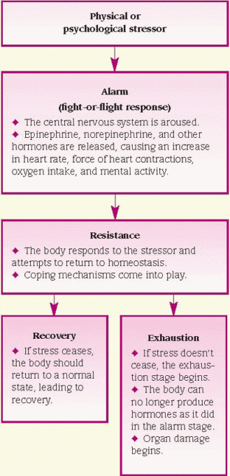
Hans Selye, a pioneer in the study of stress and disease, describes the following stages of adaptation to a stressful event: alarm, resistance, and recovery or exhaustion. (See Physical response to stress.) In the alarm stage, the body senses stress and arouses the central nervous system. The body releases chemicals to mobilize the fight-or-flight response. In this dual effort, the sympathoadrenal medullary response causes the release of epinephrine and the hypothalamic-pituitary adrenal axis causes the release of glucocorticoids. These systems work together to enable the body to respond to stressors. This release is the adrenaline rush associated with panic or aggression. In the resistance stage, the body either adapts and achieves homeostasis or it fails to adapt and enters the exhaustion stage, resulting in disease.
The stress response is controlled by actions that take place in the cells of the nervous and endocrine systems. These actions try to redirect energy to the organ that’s most affected by stress, such as the heart, lungs, or brain.
Stressful events can exacerbate some chronic diseases, such as diabetes or multiple sclerosis. Effective coping strategies can prevent or reduce the harmful effects of stress.
Cell physiology
The cell is the smallest living component of a living organism. Organisms may be made up of a single cell, such as bacteria, or billions of cells, such as human beings. In large organisms, highly specialized cells that perform an identical function are organized into tissue, such as epithelial tissue, connective tissue, nerve tissue, and muscle tissue. Tissues, in turn, form organs (such as the skin, skeleton, brain, and heart), which are integrated into body systems, such as the central nervous system (CNS), cardiovascular system, and musculoskeletal system.
CELL COMPONENTS
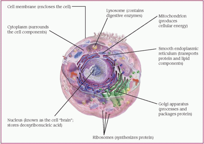
Like organisms, cells are complex units of specialized components, each component having its own function. A normal cell’s largest components are the cytoplasm, the nucleus, and the cell membrane, which surrounds the internal components and holds the cell together. (See Cell components and structures, page 4.)
Cytoplasm
The gel-like cytoplasm consists primarily of cytosol, a viscous, semitransparent fluid that’s 70% to 90% water plus various proteins, salts, and sugars. Suspended in the cytosol are many tiny structures called organelles.
Organelles are the cell’s metabolic machinery. Each performs a function to maintain the cell’s life. Organelles include mitochondria, ribosomes, endoplasmic reticulum, Golgi apparatus, lysosomes, peroxisomes, cytoskeletal elements, centrosomes, microfilaments, and microtubules.
♦ Mitochondria are spherical or rod-shaped structures that produce most of the body’s adenosine triphosphate (ATP). ATP contains high-energy phosphate chemical bonds that fuel many cellular activities. Mitochondria are the sites of cellular respiration—the metabolic use of oxygen to produce energy, carbon dioxide, and water.
♦ Ribosomes are the sites of protein synthesis.
♦ The endoplasmic reticulum is an extensive network of two varieties of membrane-enclosed tubules. The rough endoplasmic reticulum is covered with ribosomes. The smooth endoplasmic reticulum contains enzymes that synthesize lipids.
♦ The Golgi apparatus synthesizes carbohydrate molecules that combine with protein produced by the rough endoplasmic reticulum and lipids produced by the smooth endoplasmic reticulum to form such products as lipoproteins, glycoproteins, and enzymes.
♦ Lysosomes are digestive bodies that break down nutrient material as well as foreign or damaged material in cells. A membrane surrounding each lysosome separates its digestive enzymes from the rest of the cytoplasm. The enzymes digest nutrient matter brought into the cell by means of endocytosis, in which a portion of the cell membrane surrounds and engulfs matter to form a membrane-bound intracellular vesicle. The membrane of the lysosome fuses with the membrane of the vesicle surrounding the endocytosed material. The lysosomal enzymes then digest the engulfed material. Lysosomes digest the foreign matter ingested by
white blood cells by a similar process called phagocytosis.
white blood cells by a similar process called phagocytosis.
♦ Peroxisomes contain oxidases, which are enzymes that chemically reduce oxygen to hydrogen peroxide and hydrogen peroxide to water.
♦ Cytoskeletal elements form a network of protein structures that maintain the cell’s shape.
♦ Centrosomes contain centrioles, which are short cylinders adjacent to the nucleus that take part in cell division.
♦ Microfilaments and microtubules enable the movement of intracellular vesicles (allowing axons to transport neurotransmitters) and the formation of the mitotic spindle, the framework for cell division.
Nucleus
The cell’s control center is the nucleus, which plays a role in cell growth, metabolism, and reproduction. Within the nucleus, one or more nucleoli (dark-staining intranuclear structures) synthesize ribonucleic acid, a complex polynucleotide that controls protein synthesis. The nucleus also stores deoxyribonucleic acid (DNA), the double helix that carries genetic material and is responsible for cell reproduction, or division.
Cell membrane
The semipermeable cell membrane forms the cell’s external boundary, separating it from other cells and from the external environment. Roughly 75Å ( millionths of an inch) thick, the cell membrane consists of a double layer of phospholipids with protein molecules embedded in it. These protein molecules act as receptors, ion channels, or carriers for specific substances.
millionths of an inch) thick, the cell membrane consists of a double layer of phospholipids with protein molecules embedded in it. These protein molecules act as receptors, ion channels, or carriers for specific substances.Stay updated, free articles. Join our Telegram channel

Full access? Get Clinical Tree


