Immobility and Associated Problems
Learning Objectives
After studying this chapter, the student is expected to:
1. Describe the possible effect of immobility on skeletal muscle, bone, and joints.
2. Discuss the development of decubitus ulcers.
3. Explain the changes in blood pressure and potential thrombus formation.
4. List the potential problems related to respiratory function.
5. Discuss the common effects of immobility on appetite, bowel function, and urinary function.
6. Discuss the potential effects of immobility on the nervous system and psychological implications.
7. Describe the potential effect of immobility on a child’s growth.
Key Terms
atelectasis
basal metabolic rate
contracture
decubitus ulcers
diplegia
extensor
flaccidity
flexor
hemiplegia
orthostatic hypotension
osteoblastic
osteoclastic
paraplegia
pneumonia
quadriplegia
stasis
supine
Factors Involving Immobility
Immobility, or lack of movement, may involve only one part of the body such as a fractured arm in a cast. A part of the body may be affected, as occurs with paralysis: one side of the body (hemiplegia), the lower half of the body (paraplegia), or the trunk and all four limbs (quadriplegia). Diplegia refers to symmetric paralysis in any area of the body. In a coma or during an acute illness the entire body may be immobilized. The effects of such inactivity depend on the extent of the immobilization and its duration. Physiotherapy or passive exercise imposed on the involved area of the body can minimize the effects of lack of voluntary movement. Respiratory therapy is significant in preventing infections such as pneumonia.
When the body is supine (lying on the back), the loss of the force of gravity affects many of its natural functions, primarily in the intestines and urinary tract. Other noticeable effects result from the lack of stress normally exerted on bone by skeletal muscle and the decreased circulation of blood. Bed rest also alters respiratory function, metabolism, and renal function.
Musculoskeletal System Effects
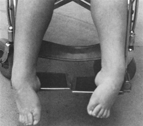
Inactive muscle loses strength, endurance, and mass very quickly. Perhaps you have seen an arm or a leg shriveled up with atrophied muscle (often called disuse atrophy) after it has been confined to a cast for several weeks. Loss of muscular strength due to immobility progresses at a rate of about 12% each week. After 3 to 5 weeks of immobility due to bed rest almost one-half of the muscular strength is lost. Correct positioning and reduction of abnormal stress on immobilized muscles and joints are important because these structures may stretch or shorten, resulting in abnormal fixation of a joint, altering biomechanics. For example, an ankle may develop contractures when a tight, heavy blanket or improper positioning puts excessive and inappropriate pressure on the foot (Fig. 25-1). Generally flexor muscles are stronger than the opposing extensor muscles (which atrophy more than the flexor muscles), and this imbalance may allow an inactive joint to take an abnormal position if flexibility is not maintained by range-of-motion exercises. With inactivity, tendons and ligaments shorten and lose elasticity. Prolonged immobility causes fibrous tissue to replace muscle cells, leading to muscle wasting and weakening, decreased flexibility, further possibly irreversible deformity (contracture), and loss of function.
The lack of muscular activity impairs venous return, which causes pooling of blood in dependent areas of the body, development of dependent edema, and a decrease in cardiac output, which may cause dizziness or fainting when changing position.
Bone deteriorates with inactivity. Bone is a “living” tissue in which new bone is constantly forming (osteoblastic activity) and other bone is being resorbed (osteoclastic activity). Bone demineralization occurs because the lack of weight bearing and muscle action reduces osteoblastic activity or bone formation; however, osteoclastic activity continues. This process leads to loss of bone mass and osteoporosis with the potential for spontaneous fractures if undue stress is placed on the bones (see Chapters 24 and 9).
The breakdown of muscle and bone tissue initially results in elevated serum levels of nitrogen wastes such as creatinine and in elevated serum calcium. Hypercalcemia may cause renal calculi or kidney stones if fluid intake is inadequate and the urine becomes too concentrated (see Chapter 18). Also a high serum calcium level can further impede muscle activity because it decreases muscle tone and leads to flaccidity or loss of muscle tone. Passive range-of-motion exercises and weight bearing if tolerated on a regular basis are helpful in preventing these complications.
Tendons and ligaments that connect the muscles to bone and maintain joint structure also require movement to maintain their structure and functionality. After 4 to 6 days of immobility these forms of connective tissue begin to shorten and the density of the tissue increases limiting flexibility and range of motion.

Cutaneous Effects
The skin breaks down easily when its circulation is impaired and cell regeneration is reduced. Blood supply is often reduced in places where the skin is stretched over bony projections and there is little fatty or muscular tissue to cushion the weight of the body. Areas that are particularly vulnerable to poor blood perfusion include the ischial tuberosities, sacrum, the greater trochanter of the hip, the heels, and the elbows. Pressure at these points causes ischemia and necrosis of tissue (Fig. 25-2), leading to decubitus ulcers (pressure sores or bedsores). Other factors that promote skin breakdown and development of decubiti include:
• Poor general circulation or anemia
• Edema
• Inadequate subcutaneous tissue in the elderly or debilitated person
• Prolonged static positioning
• Mechanical irritation or friction by clothing, braces, or other equipment
• Excessive moisture from perspiration or urine
• Inadequate nutrition or hydration
Stay updated, free articles. Join our Telegram channel

Full access? Get Clinical Tree


