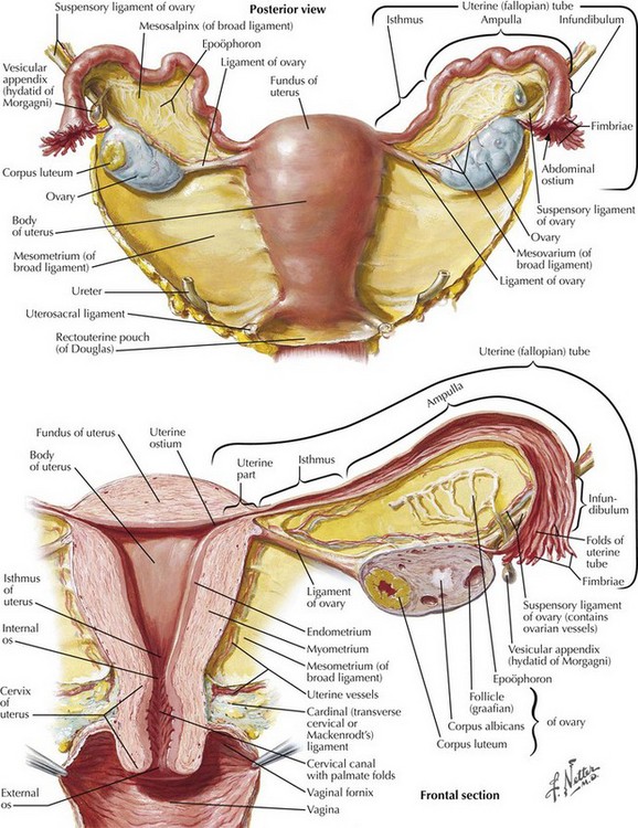
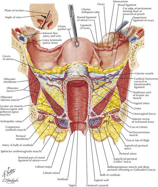
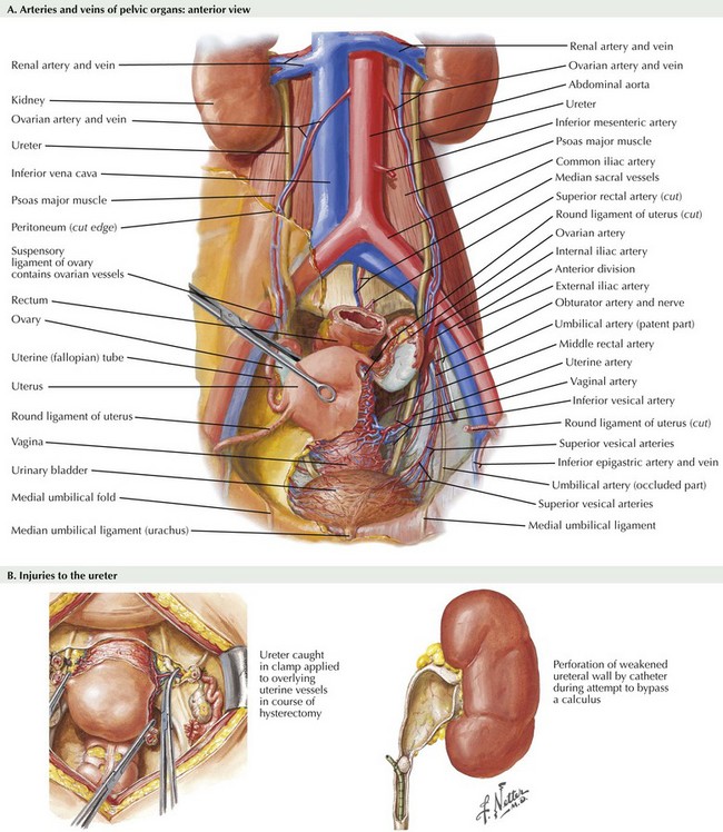
Chapter 51 Hysterectomy may be performed for both benign and malignant conditions of the uterus. The surgical approach to hysterectomy is determined by the pathology at hand as well as operator experience. Surgical approaches may include total abdominal hysterectomy, total laparoscopic hysterectomy, laparoscopic assisted vaginal hysterectomy, robotic hysterectomy or total vaginal hysterectomy. This chapter focuses on the basic description of the total abdominal extrafascial hysterectomy. Oophorectomy is described in Chapter 52. Cardinal and Uterosacral Ligaments Excellent knowledge of both intraperitoneal and extraperitoneal anatomy is critical to perform a hysterectomy. Uterine support is provided by the cardinal and uterosacral ligaments (Fig. 51-1). The cardinal ligaments extend laterally from the level of the cervical-uterine junction and divide the pelvic cavity in potential spaces: the paravesical spaces divide the cavity anteriorly and the pararectal spaces divide it posteriorly. The uterosacral ligaments extend from the cardinal ligaments posteriorly toward the ischial spines and sacrum. Between the uterosacral ligaments lies the uppermost portion of the rectovaginal septum covered by peritoneum. This area can serve as the entry point into the retrouterine space. The broad ligament consists of an anteroposterior layer of peritoneum draped over the uterus and extends from the round ligament to the infundibulopelvic ligaments posteriorly (Fig. 51-2). The retroperitoneal space and structures can be accessed through the broad ligament, which contains areolar fat. Uterine blood supply is derived from the uterine artery, which originates in the anterior branch of the hypogastric (internal iliac) artery (Fig. 51-3, A). Additional branches and collateral vessels include the vaginal and cervical branches of the uterine artery. The uterine artery crosses the lower third of the ureter before the uterine entry point at the cervicouterine junction. The majority of pelvic surgery–related ureteral injuries occur at this location, and detailed knowledge of ureteral anatomy and the relationship to the uterus and uterine blood supply is necessary to avoid iatrogenic injury to the ureter (Fig. 51-3, B).
Hysterectomy for Benign and Malignant Conditions
Introduction
Surgical Anatomy
Round and Broad Ligaments
Vascular Landmarks and Ureteral Injury
![]()
Stay updated, free articles. Join our Telegram channel

Full access? Get Clinical Tree


Hysterectomy for Benign and Malignant Conditions