Patient Story
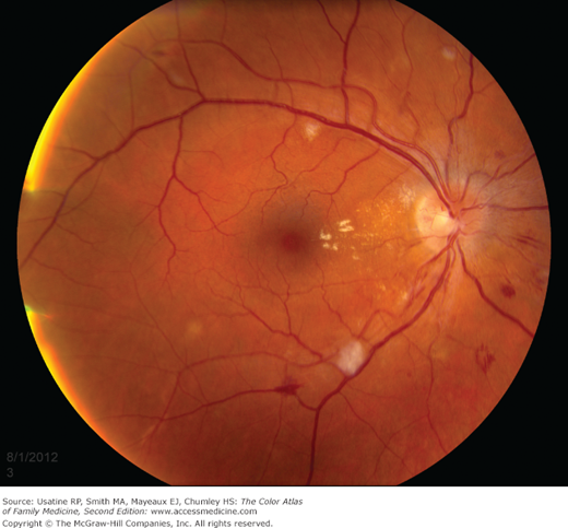
A 37-year-old man comes in for a physical examination and is noted to have a blood pressure of 198/142 mm Hg. He has no symptoms at the time. The physician performs a dilated funduscopic examination and notes optic disc edema, cotton wool spots, flame hemorrhages, dot-blot hemorrhages, arteriovenous nicking, and exudates (Figure 21-1). Fortunately, the remainder of the neurological exam and the EKG are normal. The patient is sent to the emergency room to be evaluated further and treated for a hypertensive emergency.
Introduction
Hypertensive retinopathy (HR) develops from elevated blood pressure. HR is diagnosed clinically by the presence of classic retina findings seen on funduscopic examination or digital retinal photographs in a patient with hypertension. HR can result in vision loss. Treatment is control of blood pressure.
Epidemiology
- Prevalence of 7.7% (black) versus 4.1% (white) in a population study of men and women between 49 and 73 years of age without diabetes.1
- Multiple studies show that patients with moderate HR are 2 to 3 times more likely to have a stroke than those without HR at the same level of blood pressure control independent of other risk factors.2
Etiology and Pathophysiology
- Retinal vessels become narrow and straighten at diastolic blood pressure (DBP) of 90 to 110 mm Hg.
- Arteriovenous “nicking” (white oval in Figure 21-1) occurs when the arteriolar wall enlarges from arteriosclerosis, compressing the vein. Patients with hypertension are at risk for central and branch retinal vein occlusions, which can result in significant vision loss.
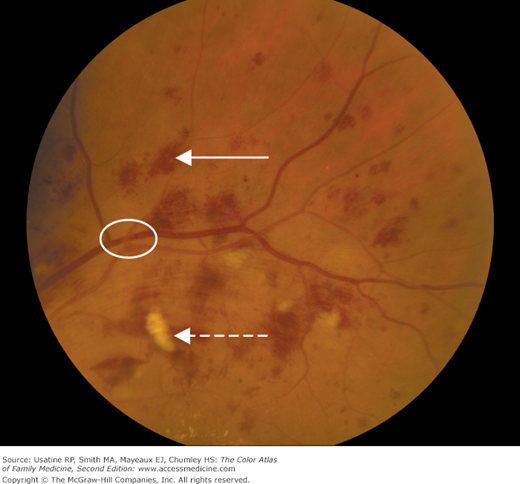
- Microaneurysms and flame hemorrhages (Figures 21-1 and 21-2) result from the increased intravascular pressure. Cotton-wool spots (dashed arrow in Figure 21-2) represent ischemia of the nerve fiber layer. Hard exudates indicate vascular leakage (white arrowheads in Chapter 20, Figure 20-1).
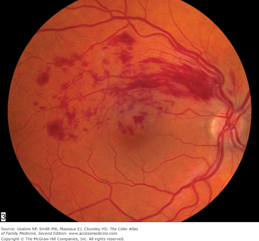
- DBP 110 to 115 mm Hg causes leakage of plasma proteins and blood products resulting in retinal hemorrhages and hard exudates (Figures 21-1, 21-2, 21-3, and 21-4).
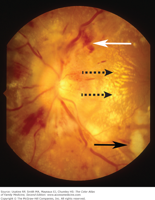
- Optic nerve swelling occurs at DBP of 130 to 140 mm Hg (Figure 21-3).
Figure 21-3
Malignant hypertensive retinopathy with optic nerve head edema (papilledema), flame hemorrhages (white arrow), cotton-wool spots (black arrow), and macular edema with exudates (dashed arrows). The patient was admitted to the hospital to treat malignant hypertension aggressively. (Courtesy of Paul D. Comeau.)


