Hyperplasia of Mesonephric Remnants
Gladell P. Paner, MD
Mahesha Vankalakunti, MD
Key Facts
Terminology
Tubular or acinar proliferation of putative mesonephric duct remnants containing characteristic intraluminal colloid-like material
Etiology/Pathogenesis
During embryogenesis, mesonephric or wolffian duct gives rise to rete testis, epididymis, vas deferens, and seminal vesicles
Mesonephric remnants may undergo proliferation
Clinical Issues
Very rare, seen in 0.6% of TURP specimens
Mean: 67 years old, range: 50-85 years
Incidental histologic finding
Typically in prostatic base but can extend into bladder neck and periprostatic soft tissues
Microscopic Pathology
Lobular or infiltrative growth of tubules that often contains dense eosinophilic colloid-like material
Lined by single layer of bland cuboidal cells with scant cytoplasm imparting “atrophic” appearance
Nuclei are regular, round, and typically with inconspicuous nucleolus but can also be prominent
Hyperplasia may be florid; tubules may appear to infiltrate between bladder neck muscle bundles and into periprostatic connective tissues
Tubules may be seen intimately associated with nerves or ganglions
Ancillary Tests
Colloid-like material is PAS(+) and PASD(+)
PSA/PAP(-)
Most are HMCK(34βE12)(+)
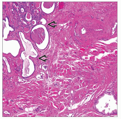 Mesonephric hyperplasia  involving prostate shows lobular and infiltrative growth of tubules, which characteristically contain intraluminal colloid-like material. involving prostate shows lobular and infiltrative growth of tubules, which characteristically contain intraluminal colloid-like material. |
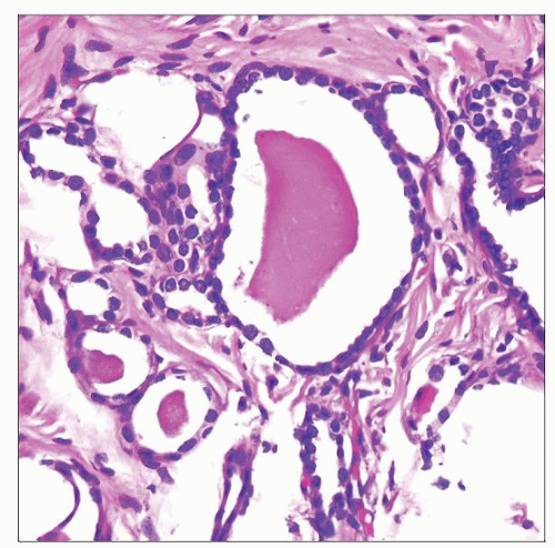 Mesonephric remnant tubules are lined by a single layer of cuboidal cells with scant cytoplasm imparting an “atrophic” appearance. Note the presence of intraluminal eosinophilic material. |
TERMINOLOGY
Definitions
Tubular or acinar proliferation of putative mesonephric duct remnants containing characteristic intraluminal colloid-like material
Histologically similar to mesonephric remnants that are well recognized in female genital tract
ETIOLOGY/PATHOGENESIS
Origin
During embryogenesis, mesonephric or wolffian duct gives rise to rete testis, epididymis, vas deferens, and seminal vesicles
Vestigial mesonephric duct remnants or rests can be seen more commonly in female genital tract and only rarely in male genital tract
Mesonephric duct remnants may undergo proliferation or hyperplasia, the cause of which is not known
CLINICAL ISSUES
Epidemiology
Incidence
Very rare
Seen in only 0.6% of transurethral resection of prostate (TURP) specimens
Only 15 cases reported in literature
Age
Mean: 67 years old, range 50-85 years
Site
Described typically in prostatic base but may extend into bladder neck and periprostatic soft tissues
Likely involves central region of prostate, since most are seen in TURP specimens
Presentation
Incidental histologic finding
Encountered mainly in patients treated for obstructive urinary symptoms due to benign prostate hyperplasia
Few cases encountered in prostatectomy specimens for prostate adenocarcinoma
Report of misdiagnosis as prostate adenocarcinoma
Treatment
None required
Prognosis


