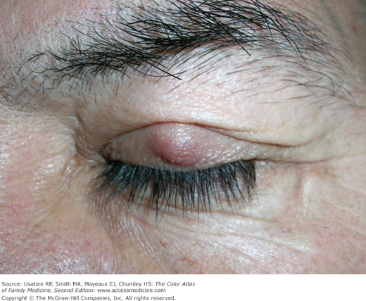
Patient Story
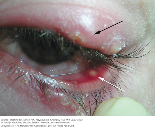
A 35-year-old woman presented with a tender nodule on the upper eyelid along with crusting and erythema to both eyelids (Figure 13-1). The upper eyelid had a large external hordeolum. When the lower eyelid was inverted, an internal hordeolum was also present. The physician recommended that she apply warm moist compresses to her eyelids 4 times a day. Her hordeola resolved within 7 days.
Introduction
Epidemiology
- Unclear incidence or prevalence in the United States, but often stated to be more common in school-age children and adults 30 to 50 years old.
- In one study of school-age children in Brazil, the prevalence of chalazion was found to be 0.2% and that of hordeolum was 0.3%.1
Etiology and Pathophysiology
- Infection in the meibomian gland (internal hordeolum), often resolves into a chalazion (Figure 13-1).
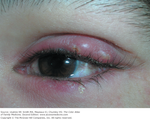
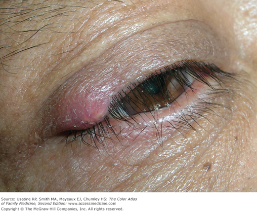
- Infection in the Zeiss or Moll gland (external hordeolum) (Figures 13-2 and 13-3).
- Staphylococcus aureus is the causative agent in most cases.
- Meibomian gland becomes blocked, often in a patient with blepharitis.
- Blocked meibomian gland’s duct releases gland contents into the soft tissue of eyelid.
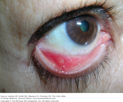
- Gland contents cause a lipogranulomatous reaction (Figure 13-4).
- Reaction can cause acute tenderness and erythema, which then resolves into a chronic nodule (Figure 13-5).
Stay updated, free articles. Join our Telegram channel

Full access? Get Clinical Tree


