Hidrocystoma (Apocrine and Eccrine)
Senait W. Dyson, MD
David Cassarino, MD, PhD
Key Facts
Terminology
Cystic, dome-shaped, translucent papules with bluish hue commonly found on the eyelids
Etiology/Pathogenesis
Most, if not all, hidrocystomas are now considered to be of apocrine origin
Clinical Issues
Relatively common
Predominately in periorbital region
Microscopic Pathology
Dermal unilocular or multilocular cyst
Often, no cyst content and appears empty
Cystic spaces lined by 1-2 layers of cuboidal or columnar epithelium
Papillary projection into cyst cavity: “Decapitation” secretion present
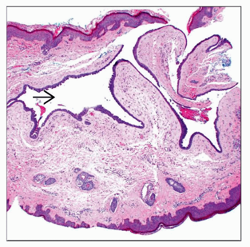 Hidrocystoma on low magnification shows characteristic thin-walled, dermal-based, empty cystic spaces  . . |
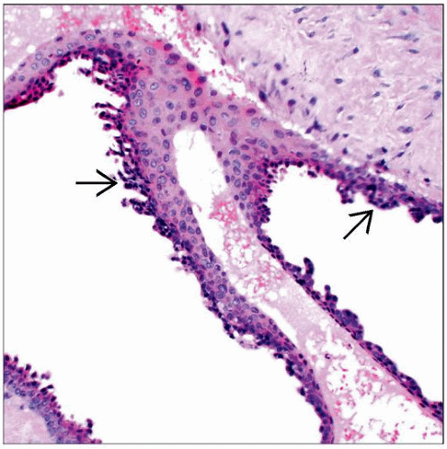 Hidrocystomas can be multilocular. A thin layer of bland-appearing apocrine epithelium  lines the cystic lumina. lines the cystic lumina. |
TERMINOLOGY
Synonyms
Cystadenoma (apocrine and eccrine)
Apocrine gland cyst
Sudoriferous cyst
Moll gland cyst
Papillary apocrine gland cyst
Definitions
Cystic, dome-shaped, translucent papules with bluish hue commonly found on the eyelids
ETIOLOGY/PATHOGENESIS
Eccrine vs. Apocrine Hidrocystomas
Most, if not all, hidrocystomas are now considered to be of apocrine origin
Apocrine hidrocystoma
Usually multiloculate
Cytoplasmic projections into cyst cavity: “Decapitation” secretion present
Eccrine hidrocystoma
Usually unilocular
No “decapitation” secretion
Considered to be retention of dilated eccrine duct or gland, and not a true cyst
Associated Disease
Schöpf-Schulz-Passarge syndrome (SSPS): Rare ectodermal dysplasia
Multiple apocrine hidrocystomas on the eyelids
Stay updated, free articles. Join our Telegram channel

Full access? Get Clinical Tree


