Patient Story
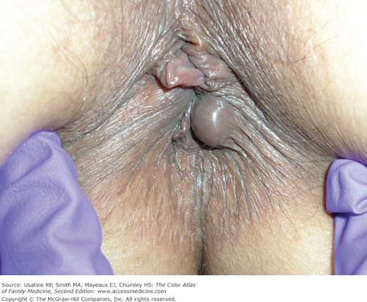
A 42-year-old woman presents to the office with rectal pressure and occasional bright red blood on the toilet paper when wiping after bowel movements (Figure 66-1). She has had difficulty with constipation off and on for many years and had large hemorrhoids during her last pregnancy. Physical examination confirms the diagnosis of external hemorrhoids.
Introduction
Epidemiology
- More than 1 million people in Western civilization suffer from hemorrhoids each year.1
- Estimated at 5% prevalence in the general population.2
- Approximately half of those older than age 50 years have experienced hemorrhoidal symptoms at some time.2
- More frequent in whites and in those of higher socioeconomic status.2
Etiology and Pathophysiology
- Three hemorrhoidal cushions (comprised of subepithelial connective tissue, elastic tissue, blood vessels, and smooth muscle) surround and support distal anastomoses between the terminal branches of the superior and middle rectal arteries and the superior, middle, and inferior rectal veins.2 The hemorrhoidal cushions have several functions, including maintaining fecal continence by engorging with blood and closing the anal canal and by protecting the anal sphincter during defecation.
- Hemorrhoidal tissue provides important sensory information, enabling the differentiation between solid, liquid, and gas and subsequent decision to evacuate.2
- Abnormal swelling of the anal cushions can occur from a number of causes (see “Risk Factors” below) resulting in increased pressure, with dilation and engorgement of the arteriovenous plexuses. Increased pressure can lead to stretching of the suspensory muscles, laxity of connective tissue, and eventual prolapse of rectal tissue through the anal canal.2 The engorged anal mucosa is easily traumatized, leading to rectal bleeding. Prolapse predisposes to incarceration and strangulation.
- Hemorrhoids are classified with respect to their position relative to the dentate line.
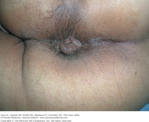
- Internal hemorrhoids (Figure 66-2) develop above the dentate line and are covered by columnar epithelium of anal mucosa. Internal hemorrhoids lack somatic sensory innervation.
- External hemorrhoids (Figure 66-1) arise distal to the dentate line. They are covered by stratified squamous epithelium and receive somatic sensory innervation from the inferior rectal nerve.
- Internal hemorrhoids (Figure 66-2) develop above the dentate line and are covered by columnar epithelium of anal mucosa. Internal hemorrhoids lack somatic sensory innervation.
- Hemorrhoids are further classified into four stages of disease severity:1,2
- Stage I—Enlargement and bleeding.
- Stage II—Protrusion of hemorrhoids with spontaneous reduction.
- Stage III—Protrusion of hemorrhoids with manual reduction possible.
- Stage IV—Irreducible protrusion of hemorrhoids usually containing both internal and external components with or without acute thrombosis or strangulation.
- Stage I—Enlargement and bleeding.
Risk Factors
Diagnosis
- Bleeding described as bright red blood (a result of the high blood oxygen content within the arteriovenous anastomoses) seen in the toilet or with wiping after bowel movements.
- Protrusion/mass (Figure 66-1).
- Pain described as a dull ache or severe if thrombosed.
- Inability to maintain personal hygiene/staining/soiling secondary to prolapse.
- Pruritus, also secondary to prolapse.
- Diagnosis is made on visual inspection and anoscopy, with and without straining:
- Physical findings of swollen blood vessels protruding from the anus (Figure 66-1).
- Excoriations may also be seen on the skin surrounding the anus.
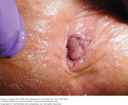
- A thrombosed hemorrhoid will be tender and firm and appear as a circular purplish bulge adjacent to the anal opening (Figure 66-3). There may be a black discoloration if there is accompanying necrosis.
- Internal hemorrhoids may be visualized on anoscopy as swollen purple blood vessels arising above the dentate line.
- Physical findings of swollen blood vessels protruding from the anus (Figure 66-1).
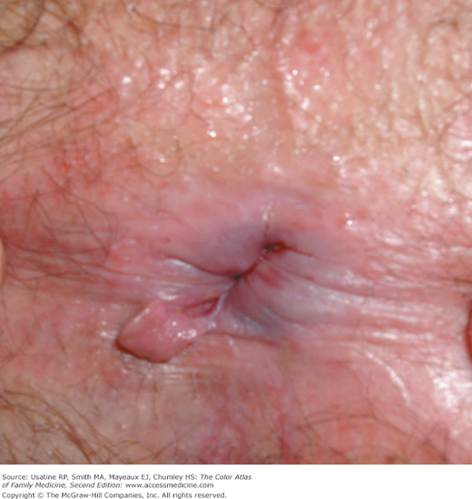
- Other physical findings that may accompany hemorrhoids are redundant tissue and skin tags (Figure 66-4) from old thrombosed external hemorrhoids.