Hemangiomas
Key Facts
Clinical Issues
Benign vascular lesions that occur in breast parenchyma and skin
May be detected as palpable masses, circumscribed masses on imaging, or incidental findings
Recurrence even after incomplete excision has not been reported
Uncommon: ˜ 1% of mastectomies or ˜ 5% of breast biopsies
Majority of vascular lesions diagnosed on core needle biopsy should undergo excision due to concern for undersampling an angiosarcoma
Microscopic Pathology
Several types of hemangioma
Perilobular hemangioma
Cavernous hemangioma
Capillary hemangioma
Complex hemangioma
Venous hemangioma
Atypical hemangiomas
Hemangiomas also occur in dermis of breast skin (nonparenchymal)
Cutaneous lesions are usually benign capillary hemangiomas
Must be distinguished from atypical vascular lesions occurring in skin after radiation treatment
Top Differential Diagnoses
Low-grade angiosarcoma
Pseudoangiomatous stromal hyperplasia
Angiolipoma
Papillary endothelial hyperplasia
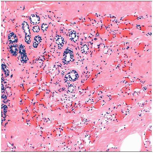
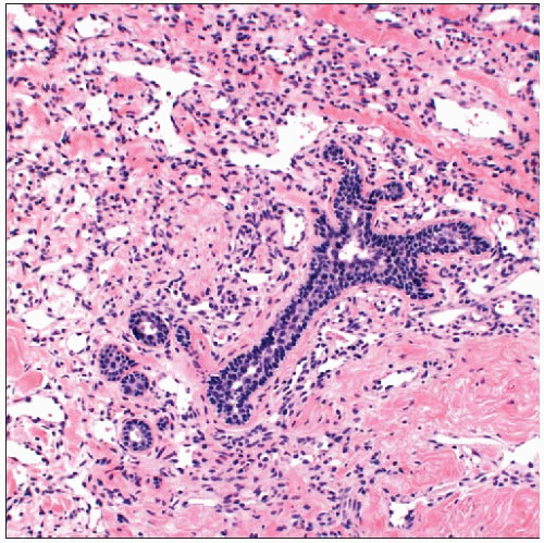
 Perilobular hemangiomas are the most common type of vascular lesion in the breast and are usually incidental findings. The small round vessels are clustered in a lobulated mass next to a lobule. |
TERMINOLOGY
Definitions
Benign lesions of vascular origin occurring in or near breast parenchyma
ETIOLOGY/PATHOGENESIS
Estrogen
Possible role for estrogen in development of hemangiomas
Hemangiomas are more common in women than in men
Hemangiomas at sites other than breast have been reported to
Occur after exogenous estrogen treatment
Increase in size during pregnancy
In a single case, a subcutaneous hemangioma of the breast was diagnosed after initiation of hormone replacement therapy
Partial involution was observed after discontinuation of hormone replacement
Hereditary Causes
Hemangiomas occur as part of several genetic syndromes
Kasabach-Merritt syndrome
Most common in extremities but can involve any anatomic site, including breast
Breast hemangiomas and angiosarcoma have been reported
Poland syndrome
Unilateral defect of pectoral muscle and ipsilateral syndactyly
Breast may be absent or hypoplastic
Rarely, congenital hemangiomas may be present
Tumors typically occur in infancy
Breast involvement is very rare
CLINICAL ISSUES
Epidemiology
Incidence
Uncommon: ˜ 1% of mastectomies or ˜ 5% of breast biopsies
May be found incidentally on microscopy of biopsy material for other indications
Age
Wide range: 18 months to 82 years
Gender
Males and females affected
Presentation
Majority are incidental findings in biopsies for other lesions
Larger hemangiomas can present as palpable mass
Some may be seen as red to purple skin lesion
May also be detected as circumscribed masses on screening mammography
Natural History
Some lesions may undergo involution or resolve spontaneously
Prognosis
Hemangiomas are benign
Recurrence after incomplete excision has not been reported
Core Needle Biopsy
Majority of vascular lesions diagnosed on core needle biopsy should undergo excision due to concern for undersampling an angiosarcoma
Vessels at periphery of an angiosarcoma can appear deceptively benign
Majority of image-detected hemangiomas are small circumscribed masses
Angiosarcomas are typically larger and have irregular borders
Changes caused by needle biopsy should not be mistaken for malignancy in excisional specimen
Hemorrhage simulating blood lakes
Thrombosis and papillary endothelial hyperplasia simulating anastomosing infiltrative vasculature
Infarction and necrosis
IMAGE FINDINGS
Mammographic Findings
Oval or lobulated mass
Punctate microcalcifications may be present
Calcifications likely related to thrombosis and phlebolith formation (coarse or eggshell-like calcifications)
Ultrasonographic Findings
Oval mass with circumscribed margins
MACROSCOPIC FEATURES
General Features
Hemangiomas are well-circumscribed, soft, spongy masses
Rarely palpable
Reddish brown color
Size
Typically small (0.5-2 cm)
Lesions > 2 cm are more likely to be angiosarcomas
MICROSCOPIC PATHOLOGY
Histologic Features
Perilobular hemangioma
Most common vascular breast lesion (1-5% of breast biopsies)
Typically, small incidental microscopic finding
Often intimately intermingled with lobular and terminal duct epithelial structures but can be located anywhere in breast tissue
Vascular spaces are lined by flattened endothelium
Vessels are small and rounded with little or no muscular coat
Atypical perilobular hemangiomas have hyperchromatic nuclei and may have anastomosing vascular channels
Cavernous hemangioma
Composed of large cavernous vascular channels
Dilated thin-walled vessels are lined by flattened endothelium, congested with blood
Vessels appear separate; anastomosing pattern would be unusual
Smaller vessels may be present focally or at periphery
Thrombosis may be present with papillary endothelial hyperplasia (Masson lesion)
Stay updated, free articles. Join our Telegram channel

Full access? Get Clinical Tree


