Incidence ranges from < 1% to 7.3% in autopsy studies
Clinical Issues
• Majority discovered incidentally
Clinically silent
– Tumors under 4 cm rarely symptomatic
When symptomatic, present with abdominal pain, hepatomegaly, palpable mass
• More frequent in older patients and women
• Treatment is surgical resection or ablative therapy if symptomatic; otherwise observation
• Complications rare but include rupture and consumptive coagulopathy
Macroscopic
• Usually solitary and subcapsular
• Cut surface shows dark red, spongy mass composed of blood-filled cavities
• Most < 4 cm
Microscopic
• Dilated, variably sized vascular spaces
Lined by flat, bland endothelial cells
Fibrin thrombi may be present in vascular spaces
• Connective tissue septa of varying widths
• Older lesions frequently contain involutional changes such as fibrosis, thrombosis, calcification
Can usually detect underlying vascular architecture even if involutional change is extensive
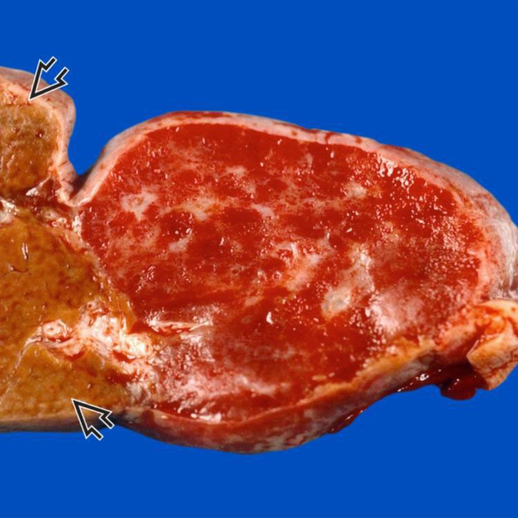
Gross Specimen The cut surface of this partial hepatectomy specimen shows a spongy red mass directly beneath the capsule. Normal liver is to the left of the tumor . (Courtesy G. Gray, MD.)
Dilated Vascular Channels Filled With Blood This hemangioma has a somewhat irregular interface with the normal liver on the right. The lesion consists of dilated vascular channels filled with blood with intervening fibrous septa.
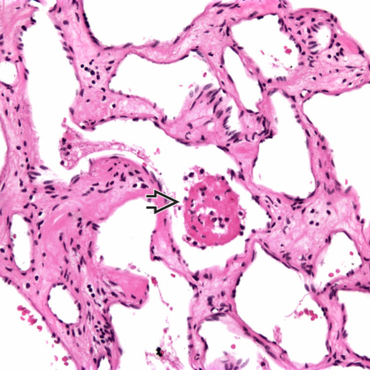
Dilated Vascular Spaces Hemangiomas are composed of dilated vascular spaces with a bland, flat endothelial lining. The intervening fibrous bands are paucicellular and of varying thickness. Note the organizing thrombus in a vascular space .
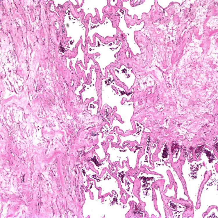
Fibrosis This hemangioma shows marked fibrosis, a common involutional change due to thrombosis over time. Residual typical dilated vascular channels are seen in the center of the picture.
TERMINOLOGY
Synonyms
• Cavernous hemangioma, sclerosing hemangioma
Definitions
• Benign vascular tumor
Most common primary tumor of liver
ETIOLOGY/PATHOGENESIS
Unknown
• Possibly congenital
• Postulated but unproven role of sex hormones
CLINICAL ISSUES
Epidemiology
• Incidence
Ranges from < 1% to 7.3% in autopsy studies
Only gold members can continue reading. Log In or Register to continue


 . (Courtesy G. Gray, MD.)
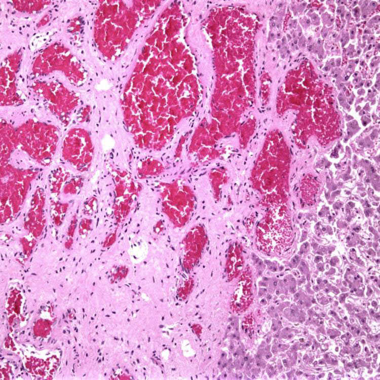
 .
.