QUESTION 29.1
A. Hereditary
B. Idiopathic
C. Infectious
D. Metabolic
E. Toxic
2. A 24-year-old male suffers from congestive heart failure. He has no history of therapeutic or illicit drug use, valvular disease, or systemic disorder. A myocardial biopsy shows myofibers arranged as seen in this picture. Occasional fibers contain basophilic granules. These morphologic features are most consistent with:
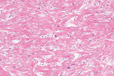
QUESTION 29.2
A. Amyloidosis
B. Dilated cardiomyopathy
C. Hypertrophic cardiomyopathy
D. Infiltrative cardiomyopathy
E. Myocarditis
3. A 30-year-old woman presents with skin rash and signs and symptoms of congestive heart failure. Investigations reveal impaired diastolic function with preserved systolic function. An endomyocardial biopsy shows thickened endocardium covered with thrombus, as seen in this picture. A mononuclear and eosinophilic infiltrate is seen in the endocardium. Which of the following is the most likely diagnosis?
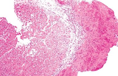
QUESTION 29.3
A. Acute rheumatic fever
B. Arrhythmogenic right ventricular cardiomy- opathy
C. Endocardial fibroelastosis
D. Idiopathic restrictive cardiomyopathy
E. Loeffler endocarditis
4. An endomyocardial biopsy reveals expansion of the myocardial interstitium by a pale eosinophilic substance. Congo red staining is positive. Deposition of which of the following is the most frequent cause of this condition?
A. AA type (inflammatory)
B. AL type (immunoglobulin associated)
C. Atrial natriuretic factor
D. Beta-2 microglobulin
E. Transthyretin protein
5. A patient treated with chemotherapy for a lymphoid malignancy undergoes an endomyocardial biopsy, which is fixed in glutaraldehyde for ultrastructural analysis. The light microscopic appearance of toluidine-stained thick sections and the changes detected by electron microscopy (EM) are shown in these photomicrographs. Which of the following is the most likely cause of these findings?
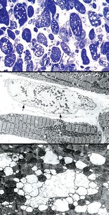
QUESTION 29.5
A. Anthracycline toxicity
B. Busulfan toxicity
C. Chloroquine toxicity
D. Cyclophosphamide toxicity
E. Fabry disease
6. In order to make a morphologic diagnosis of definite myocarditis, which of the following changes is required?
A. Eosinophilic infiltration
B. Giant cells
C. Inflammatory infiltration
D. Inflammatory infiltration and fibrosis
E. Inflammatory infiltration and myocyte necrosis
7. A 27-year-old man presents with rapid onset of heart failure and ventricular arrhythmias. An endomyocardial biopsy reveals the changes shown in this picture. Which of the following is the most likely diagnosis?
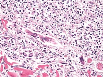
QUESTION 29.7
A. Giant cell myocarditis
B. Hypersensitivity myocarditis
C. Lymphocytic myocarditis
D. Sarcoidosis
E. Trypanosomiasis
8. According to the 2004 revised grading system of the International Society for Heart and Lung Transplantation (ISHLT), an endomyocardial biopsy with a single infiltrate of mononuclear infiltrates distorting the myocardial architecture and associated with myocyte damage is graded as:
A. 0 R
B. 1 R
C. 2 R
D. 3 R
9. Two weeks after cardiac transplantation, an endomyocardial biopsy demonstrates a single well-demarcated, mononuclear infiltrate in a subendocardial location, as shown in this photomicrograph. Numerous plasma cells are found within the aggregate. Immunostains reveal CD20-positive B lymphocytes and CD21-positive dendritic cells in the core of the lesion, surrounded by a rim of CD3-positive T cells. Which of the following is the most likely morphologic diagnosis?
QUESTION 29.9
A. Acute rejection, grade 2
B. Cyclosporin A toxicity
C. Nodular endocardial infiltrate (Quilty effect)
D. Posttransplantation lymphoproliferative disorder
E. Prior biopsy site
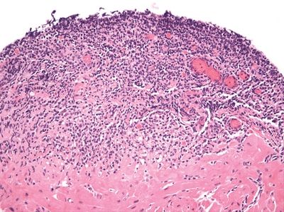
10. An endomyocardial biopsy of a heart allograft reveals interstitial edema, sparse lymphohistiocytic infiltrates, and swelling of capillary endothelium. The key findings of immunohistochemistry for CD68 and complement component C4d are shown in these photomicrographs. These findings are typically associated with:
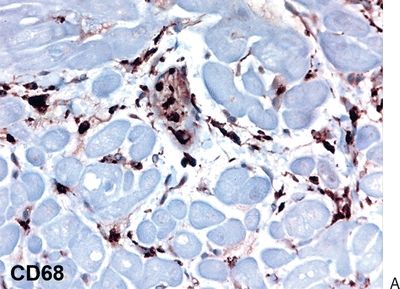
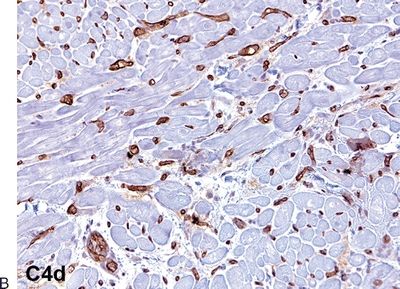
QUESTION 29.10
Stay updated, free articles. Join our Telegram channel

Full access? Get Clinical Tree


