 Chapter 2 Head
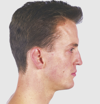
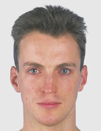
Face (Figs 2.1–2.7)
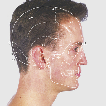
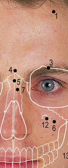
Facial bones (Figs. 2.3, 2.4)
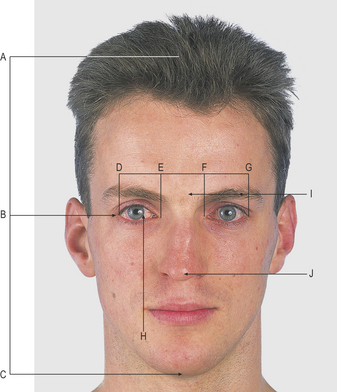
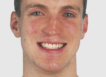
2.2 Facial alignment
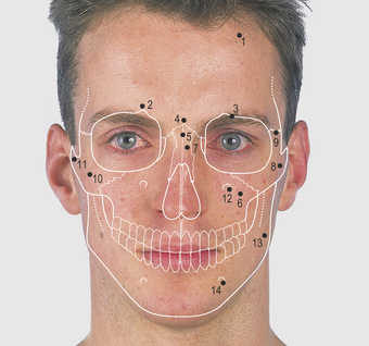

The two nasal bones articulate with each other, with the frontal bone and with the frontal process of the maxillary bone. The internasal and frontonasal sutures meet at the nasion. The lateral border of the orbit is formed by the frontal and zygomatic bones and the frontozygomatic suture can be palpated along this margin. The zygomatic bone forms the prominence of the cheek and, with the maxillary bone, the inferior margin of the orbit. It has a posterior process which, with the zygomatic process of the temporal bone, forms the zygomatic arch (Fig. 2.17).
The inferior alveolar margin of each maxilla carries the sockets for the teeth and the bone houses the maxillary air sinus. It also forms parts of the lateral wall of the nose and the hard palate (Fig. 2.25). The infraorbital foramen in the maxilla is in line with the supraorbital notch; it is 1cm below the inferior orbital margin and transmits the infraorbital nerve.
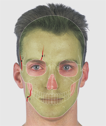
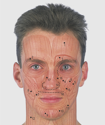
Facial muscles (Fig. 2.6)
The skin around the face is thin, vascular, sensitive and, in the male, hairy with abundant sweat and sebaceous glands. There is a variable amount of fat but no deep fascia. The skin over the nose is adherent to the nasal cartilages but not to the nasal, frontal or maxillary bones. The two nasal cavities are separated by the midline nasal septum and open anteriorly at the anterior nares. Examination of these openings using a torch reveals the wider vestibule within the expanded ala and usually the anterior end of the inferior turbinate bone. The muscles of facial expression are arranged as sphincters and dilators around the orbit, nose and mouth (Fig. 2.6). Their actions are linked with the mood of an individual and they are innervated by the facial (7th cranial) nerve (Fig. 2.13). The frontalis muscle blends with the occipital aponeurosis, and the platysma with the subcutaneous tissue of the neck and upper thorax.
In deep cuts of the face, sutures are placed in the muscles as well as the skin. Incisions follow skin creases, such as around the side and base of the nose and along the medial aspect of the orbit, or are mucosal within a cavity such as the mouth. The three incisions shown in Figure 2.6 can all be used to gain access to the sphenoidal air sinus and the pituitary gland in spite of the depth of the latter structure. A microscope, a strong light and long fine instruments are used.
Lateral aspect of the head (Figs 2.8–2.18)
The lateral aspect of the skull vault is formed of the frontal, parietal and occipital bones; the first two meet in the midline at the bregma and the last two at the lambda (Fig. 2.9). These sites are unfused at birth and are known respectively as the anterior and posterior fontanelles. The triangular-shaped posterior fontanelle (lambda) closes 2–3 months after birth and the diamond-shaped anterior (bregma) at approximately 18 months. Four bones (frontal, parietal, greater wing of sphenoid and the squamous temporal bone) meet in H-shaped fashion in the temporal region, overlying the middle meningeal artery; this point is known as the pterion. A burr hole is placed through the skull at the pterion to evacuate the blood clot when there is uncontrolled haemorrhage from a damaged middle meningeal artery, as in a skull fracture. (The pterion is also called the sylvian point because it overlies the stem of the sylvian, or lateral, cerebral sulcus.)