 Chapter 4 Thorax
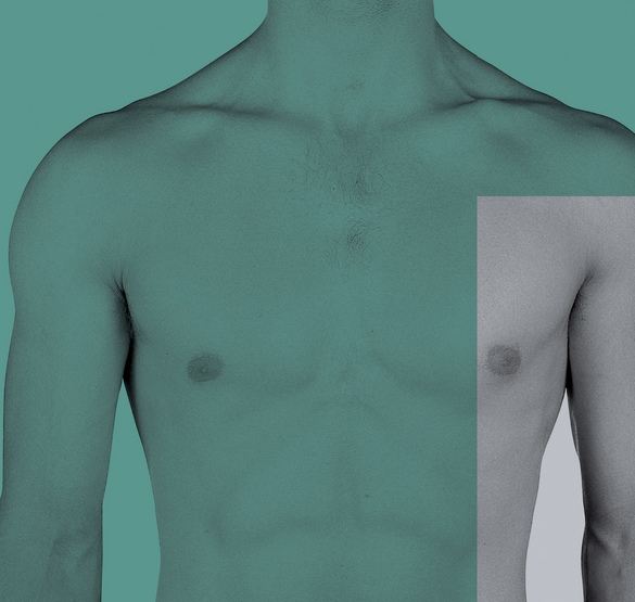
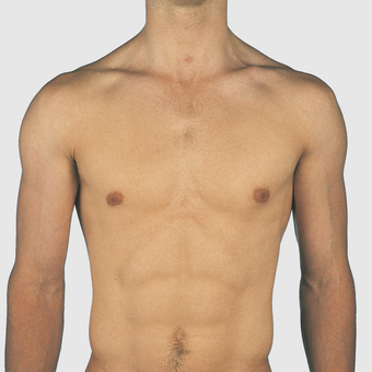
Anterior chest wall (Figs 4.1–4.6)
The chest extends from the clavicles above to the inferior costal margin below. It is formed of the ribs and costal cartilages, the sternum and associated muscles; the two sides of the chest are usually symmetrical. The rate, depth and character of respiration can be observed, as can the apex beat of the heart on the left side. The midline sternum is made up of the manubrium, the body and the xiphisternum from above downwards (Fig 4.2). The suprasternal notch on the superior aspect of the manubrium is palpable. The manubrium and body are also palpable throughout their length and they are united by a secondary cartilaginous joint forming the sternal angle (the angle of Louis). The angle is at the level of the lower border of the fourth thoracic vertebral body. It forms an important landmark for the description of structures inside the chest. Ribs are counted from this site, as it is consistently palpable: the second costal cartilage articulates on each side of the manubriosternal joint. The xiphisternum is covered by the rectus abdominis muscles and is less easily palpable. It is of variable length and when long, and suddenly noted by a subject, may be thought to be abnormal. The xiphisternal joint is at the level of the ninth thoracic vertebral body.
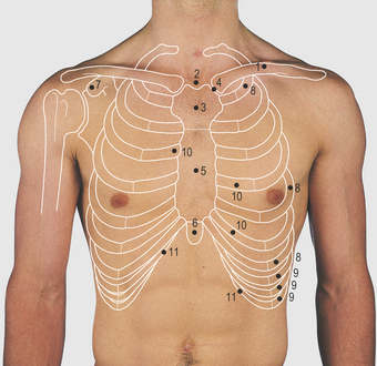
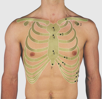
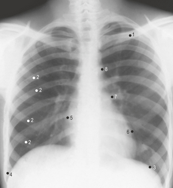
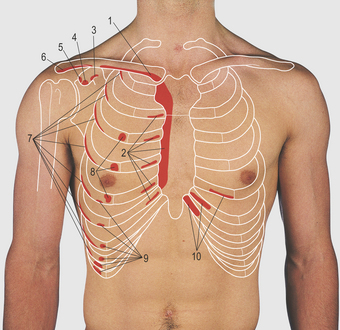
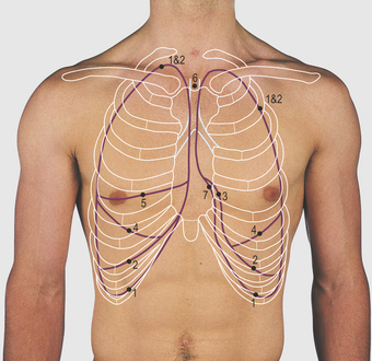
The rib cage is made up of 12 pairs of ribs, each having a posterolateral bony and an anterior costal cartilaginous component (Fig 4.2). In a thin male subject, many ribs are visible but they may be obscured by overlying muscle, fat or breast tissue. These structures can also make it difficult to count ribs by palpation. The first rib is not easily palpated, being deep to the fibres of the pectoralis major muscle and the clavicle. The second is consistently palpable at its cartilaginous articulation with the manubriosternal junction. The upper seven (true) ribs articulate directly with the sternum via their costal cartilages, whereas the eighth to 10th (false) ribs articulate via their costal cartilages with the cartilage of the rib above. The 11th and 12th (floating) ribs are considered in Figure 4.9 (p. 35). The lower costal margin is formed by the lower six ribs and their costal cartilages. The number of the ribs and intercostal spaces are used when describing normal and abnormal findings of the chest wall or thoracic cavity. The intercostal spaces are filled by the intercostal muscles, attached to the adjacent ribs.
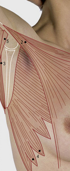
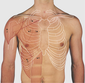
The clavicle is palpable throughout its length; it articulates medially at the sternoclavicular joint. Like the temporomandibular joint, it has a fibrocartilaginous covering of its articular surfaces and contains a fibrocartilaginous disc. The pectoralis major muscle is attached medially to the clavicle and upper five to seven costal cartilages, their related half of the sternum, and from the sheath of the rectus abdominis (Fig 4.6). Its fibres pass laterally to the lateral lip of the bicipital groove of the humerus and form the bulk of the anterior axillary fold (Fig 4.14
Stay updated, free articles. Join our Telegram channel

Full access? Get Clinical Tree


