Back

image image Chapter 6 Back


The axial skeleton is made up of the skull (pp. 10 and 13), the spine and the rib cage (p. 32–34). The spine (vertebral column) is an osseocartilaginous structure lying posteriorly in the midline. It articulates with the skull superiorly and extends through the neck and trunk to the tip of the coccyx. It articulates with the rib cage, forms part of the pelvis and gives attachment to the muscles of the shoulder girdle. The column thus provides a rigid, yet flexible, axis for the head to pivot; the upper limbs to be suspended and carry loads; the thoracic cage to be attached and function as an expansile respiratory unit; and the transfer of the body weight to the legs, when standing and in locomotion, and to the ischial tuberosities when seated. The column also provides a protective covering for the spinal cord.


The vertebral column is made up of 33 vertebrae – seven cervical, 12 thoracic, five lumbar, five sacral and four coccygeal. Of these, 24 are mobile: the sacral vertebrae are fused and the coccygeal vestigial. The mobile vertebrae are united by fibrocartilaginous intervertebral discs, each comprising an outer firm ring, the annulus fibrosus, and a central gelatinous nucleus pulposus. The discs make up a quarter of the height of the column.


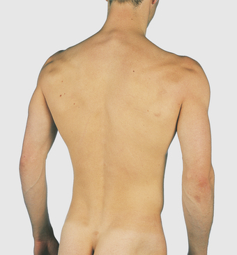
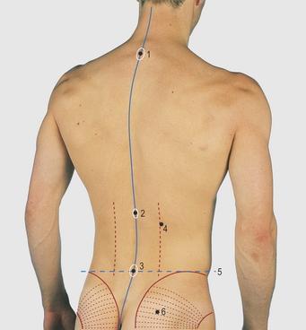
The embryonic and early fetal vertebral column is curved, concave ventrally (flexed) throughout its length – the primary curvature. Two secondary curvatures, concave dorsally, develop in the cervical and lumbar regions. The cervical curve starts as early as the 10th week ‘in utero’. After birth, further extension in the cervical region is produced by the muscles raising the head, and extension in the lumbar region accompanies the adoption of the erect posture. The thoracic and sacral regions retain the primary curvature (Figs 6.1 and 6.2).


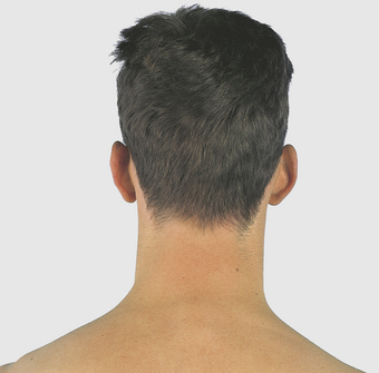
Posterior aspect of the neck


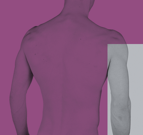
The posterior aspect of the neck is bounded superiorly by the superior nuchal line of the occipital bone extending medially to the external occipital protuberance and laterally to the mastoid process (Figs 6.36.6).


The structures in the lower part of the neck are less easily discernible posteriorly as they are covered by the trapezius, splenius and powerful erector spinae muscles. The ligamentum nuchae lies in the midline between the muscles of the two sides, and is attached to the occipital bone and the cervical spines. The spines of the lower two or three cervical, and all the thoracic, vertebrae are palpable. The upper cervical spines are impalpable and overlain by a median furrow. The non-bifed lowest (seventh) cervical vertebral spine and the first thoracic are the most obvious, the former being termed the vertebra prominens.


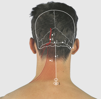
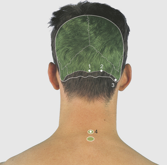
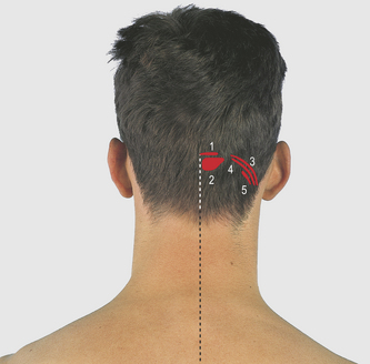
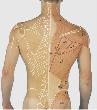
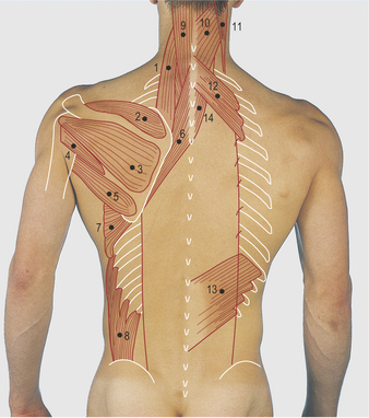
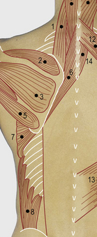
The trapezius muscle has a wide attachment (Figs 6.7 and 6.8, see also p. 70). It lies superficially and is attached superiorly to the medial third of the superior nuchal line, to the ligament nuchae and the spines and interspinous ligaments of the cervical and thoracic vertebrae. Laterally, it is attached to the acromion and spine of the scapula, both of which are palpable from the tip of the shoulder to the medial angle of the bone. The sternocleidomastoid muscle is attached to the lateral third of the superior nuchal line and to the mastoid process. The occipital artery emerges medial to the muscle to pass over the skull; it is palpable at this site.


Jun 16, 2016 | Posted by in ANATOMY | Comments Off on Back

Full access? Get Clinical Tree

Get Clinical Tree app for offline access