Genital Tract Infections


Genital Tract Infections


General Considerations


Anatomy


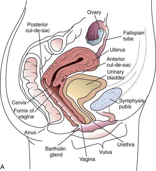
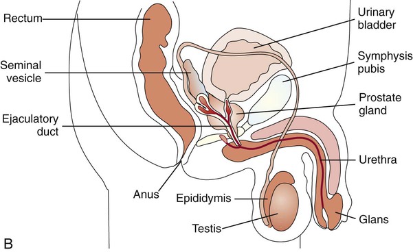
Familiarity with the anatomic structures is important for appropriate processing of specimens from genital tract sites and interpretation of microbiologic laboratory results. The key anatomic structures for the female and male genital tract in relation to other important structures are shown in Figure 74-1.


The female reproductive system consists of two main parts: the uterus and the ovaries. The uterus produces vaginal and uterine secretions and is the location where the human fetus grows and matures during reproduction. The ovaries connect to the uterus and the fallopian tubes. The ovary produces the female eggs that pass through the fallopian tubes and will imbed in the uterus when fertilized by the male sperm. The uterus connects to the vaginal opening through the cervix.


The male reproductive system, unlike the female, consists of a number of organs that are located external to the abdominal cavity. The main organs consist of the penis and the testis that produce the semen and sperm for fertilization of the female egg. The sperm is stored in a small gland coiled around the testis, the epididymis. The prostate gland surrounds the ejaculatory duct and produces semen, prostatic fluid, and seminal fluid.


Resident Microbial Flora


The lining of the human genital tract consists of a mucosal layer of transitional, columnar, and squamous epithelial cells. Various species of commensal bacteria colonize these surfaces, causing no harm to the host except under abnormal circumstances. The colonization of the surface by resident flora produces a biologic barrier preventing the adherence of pathogenic organisms. Normal urethral flora includes coagulase-negative staphylococci and corynebacteria, as well as various anaerobes. The vulva and penis, especially the area underneath the prepuce (foreskin) of the uncircumcised male, may harbor Mycobacterium smegmatis along with other gram-positive bacteria.


The flora of the female genital tract varies with the pH and estrogen concentration of the mucosa, which depends on the host’s age. Prepubescent and postmenopausal women harbor primarily staphylococci and corynebacteria (the same flora present on surface epithelium), whereas women of reproductive age may harbor large numbers of facultative bacteria such as Enterobacteriaceae, streptococci, and staphylococci, as well as anaerobes such as lactobacilli, anaerobic non-spore-forming bacilli and cocci, and clostridia. Lactobacilli are the predominant organisms in secretions from normal, healthy vaginas. Recent studies have shown that hydrogen peroxide–producing lactobacilli are associated with a healthy state. The numbers of anaerobic organisms remain constant throughout the monthly cycle. Many women carry group B beta-hemolytic streptococci (Streptococcus agalactiae), which may be transmitted to the neonate. Although yeasts (acquired from the gastrointestinal tract) may be transiently recovered from the female vaginal tract, they are not considered normal flora.


Sexually Transmitted Diseases and Other Genital Tract Infections


Genital tract infections may be classified as endogenous or exogenous. Exogenous infections may be acquired as people engage in sexual activity, and these infections are referred to as sexually transmitted diseases (STDs). In contrast, endogenous infections result from normal genital flora.


In the female, genital tract infections can be divided between lower genital tract (vulva, vagina, and cervix) and upper genital tract (uterus, fallopian tubes, ovaries, and abdominal cavity) infections. Lower genital tract infections are commonly acquired by sexual or direct contact. Although the organisms that cause lower genital tract infections are not usually part of the normal genital tract flora, some organisms normally present in very low numbers can increase sufficiently to cause disease. Upper genital tract infections are frequently an extension of a lower tract infection in which organisms from the vagina or cervix travel into the uterine cavity and on through the endometrium to the fallopian tubes and ovaries. Similarly, an organism can spread along contiguous mucosal surfaces in the male from a lower genital tract site of infection (i.e., urethra) and cause infection in a reproductive organ such as the epididymis.


Genital Tract Infections


Sexually Transmitted Diseases and Other Lower Genital Tract Infections


Lower genital tract infections may be acquired either through sexual contact with an infected partner or through nonsexual means. These infections are some of the most common infectious diseases.


Epidemiology/Etiologic Agents


STDs are major public health problems in all populations and socioeconomic groups worldwide. An estimated 448 million new cases of curable STDs occur each year worldwide. The incidence and spread of STDs are greatly influenced by numerous factors such as the availability of multiple sexual partners, the presence of asymptomatic infection, the frequent movement of people within populations, and increasing affluence.


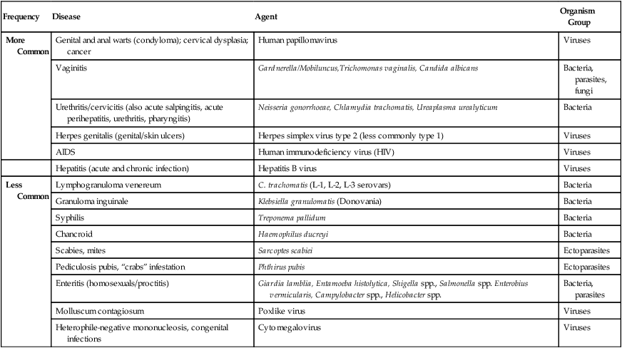
The number of microorganisms that can cause genital tract infections is large. These organisms are diverse, representing all four major groups of microorganisms (bacteria, viruses, fungi, and parasites). The major causes of genital tract infections are listed in Table 74-1.


Routes of Transmission


Although genital tract infections can be caused by members of the patient’s genital flora (endogenous infections), the overwhelming majority of lower genital tract infections are sexually transmitted.


Sexually Transmitted.

Chlamydia trachomatis, Neisseria gonorrhoeae, Trichomonas vaginalis, human immunodeficiency virus (HIV), Treponema pallidum, Ureaplasma urealyticum, Mycoplasma hominis, other mycoplasmas, herpes simplex virus (HSV), and others may be acquired during sexual activity. In addition, other agents that cause genital tract disease and may be sexually transmitted include adenovirus, coxsackievirus, molluscum contagiosum virus (a member of the poxvirus group), the human papillomaviruses (HPVs) of genital warts (condylomata acuminata; types 6, 11, and others), and those associated with cervical carcinoma (predominantly types 16 and 18, but numerous others are also implicated), Klebsiella granulomatis, and ectoparasites such as scabies and lice. Some of these agents are not routinely isolated from clinical specimens. Infections with more than one agent may occur; therefore, dual or concurrent infections should always be considered.


An individual’s sexual habits and practices dictate potential sites of infection. Homosexual practices and increasingly common heterosexual practices of anal-genital or oral-genital intercourse allow for transmission of a genital tract infection to other body sites such as the pharynx or anorectic region. In addition, these practices have required that other gastrointestinal and systemic pathogens also be considered etiologic agents of STDs. The intestinal protozoa Giardia lamblia, Entamoeba histolytica, and Cryptosporidium spp. are significant causes of STDs, especially among homosexual populations. In the same group of patients, fecal pathogens, such as Salmonella, Shigella, Campylobacter, and Microsporidium, are often transmitted sexually. Oral-genital practices may provide an opportunity for N. meningitidis to colonize and infect the genital tract. Viruses shed in secretions or present in blood (cytomegalovirus [CMV]; hepatitis B, possibly C and E; other non-A, non-B hepatitis viruses; human T-cell lymphotropic virus type I [HTLV-I]; and HIV) are spread by sexual practices.


Certain infections that are sexually transmitted occur on the surface epithelium of or near the lower genital tract. The major pathogens of these types of infections include HSV, Haemophilus ducreyi, and T. pallidum.


Other Routes.

Organisms may also be introduced into the genital tract by instrumentation, presence of a foreign body, or irritation and can subsequently cause infection. These infections are often a result of infection with the same organisms capable of causing skin or wound infections. Infection can also be transmitted from mother to infant either in vivo (within the living body) or during delivery. For example, transplacental infection may occur with syphilis, HIV, CMV, or HSV. Infection in the newborn can also be acquired during delivery by direct contact with an infectious lesion or discharge in the mother and a susceptible mucous membrane such as the eye in the infant. STDs, such as HSV, C. trachomatis, and N. gonorrhoeae, may be transmitted from mother to newborn in this manner. Other organisms, such as group B streptococci, Escherichia coli, and Listeria monocytogenes originating from the mother may also be transmitted to the infant before, during, or after birth. (Infections in the fetus and newborn are discussed later in this chapter.)


Clinical Manifestations


Clinical manifestations of lower genital tract infections are as varied and diverse as the etiologies.


Asymptomatic.

Although symptoms of genital tract infections generally cause the patient to seek medical attention, a patient with an STD, especially a female, may be free of symptoms (i.e., asymptomatic). For example, gonorrhea (Neisseria gonorrhoeae) or chlamydia (Chlamydia trachomatis) infection in the male is usually obvious because of a urethral discharge, yet females with either or both of these infections may have either minimal symptoms or no symptoms at all. Also, the primary lesion of syphilis (chancre) can be unremarkable and go unnoticed by the patient. Therefore, the lack of symptoms does not guarantee the absence of disease. Unfortunately, these asymptomatic individuals can serve as reservoirs for infection and unknowingly spread the pathogen to other individuals. Asymptomatic infections in the female caused by N. gonorrhoeae or C. trachomatis that go untreated can lead to serious sequelae such as pelvic inflammatory disease or infertility.


Lesions of the Skin and Mucous Membranes.

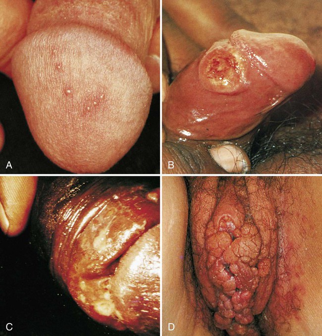
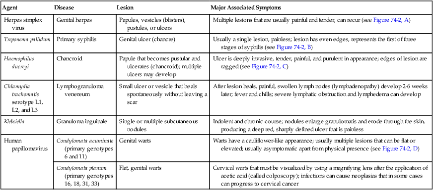
Numerous organisms can cause genital lesions that are diverse in both their appearance and their associated symptoms (Figure 74-2). The agents and their features of infection are summarized in Table 74-2. Some of these infections, such as genital herpes (caused by HSV) or genital warts (caused by HPVs and discussed in Chapter 66), are common, whereas others, such as lymphogranuloma venereum and granuloma inguinale, are uncommon in the United States. Specific HPV genotypes infect mucosal cells in the cervix and can cause a progressive spectrum of abnormalities classified as low-grade and high-grade squamous intraepithelial neoplasia (process of rapid cell growth that is faster than normal and continues to grow, i.e., a tumor) and in some cases, progress to invasive cervical cancer.


TABLE 74-2


Summary of Common Causes of Genital Lesions of the Skin and Mucous Membranes


Agent Disease Lesion Major Associated Symptoms
Herpes simplex virus Genital herpes Papules, vesicles (blisters), pustules, or ulcers Multiple lesions that are usually painful and tender, can recur (see Figure 74-2, A)
Treponema pallidum Primary syphilis Genital ulcer (chancre) Usually a single lesion, painless; lesion has even edges, represents the first of three stages of syphilis (see Figure 74-2, B)
Haemophilus ducreyi Chancroid Papule that becomes pustular and ulcerates (chancroid); multiple ulcers may develop Ulcer is deeply invasive, tender, painful, and purulent in appearance; edges of lesion are ragged (see Figure 74-2, C)
Chlamydia trachomatis serotype L1, L2, and L3 Lymphogranuloma venereum Small ulcer or vesicle that heals spontaneously without leaving a scar After lesion heals, painful, swollen lymph nodes (lymphadenopathy) develop 2-6 weeks later; fever and chills; severe lymphatic obstruction and lymphedema can develop
Klebsiella Granuloma inguinale Single or multiple subcutaneous nodules Indolent and chronic course; nodules enlarge granulomatis and erode through the skin, producing a deep red, sharply defined ulcer that is painless
Human papillomavirus Condylomata acuminate (primary genotypes 6 and 11) Genital warts Warts have a cauliflower-like appearance; usually multiple lesions that can be flat or elevated; usually asymptomatic apart from physical presence (see Figure 74-2, D)
Condylomata planum (primary genotypes 16, 18, 31, 33) Flat, genital warts Cervical warts that must be visualized by using a magnifying lens after the application of acetic acid (called colposcopy); infections can cause neoplasias that in some cases can progress to cervical cancer

image


Vaginitis.

Inflammation of the vaginal mucosa, called vaginitis, is a common clinical syndrome accounting for approximately 10 million office visits each year. Women who present with vaginal symptoms often complain of an abnormal discharge and additional symptoms such as an offensive odor or itching. Vulvitis, local irritation of external genitalia, may be associated with vaginitis. The three most common causes of vaginitis in premenopausal women are vaginal candidiasis, bacterial vaginosis (group B streptococci, E. coli, and enterococci), and trichomoniasis.


Candida albicans causes about 80% to 90% of cases of vaginal candidiasis; other species of Candida account for the remaining cases. Yeast can be carried vaginally in small numbers and produce no symptoms. Most patients experiencing candidiasis complain of perivaginal itching, often with little or no discharge. Irritating symptoms such as erythema are also associated with candidiasis. Frequently, discharge is classically thick and “cheesy” in appearance.


Vaginal infection with T. vaginalis, a protozoan parasite, produces a profuse, slightly offensive, yellow-green discharge; patients frequently complain of itching. About 25% of women carrying trichomonads are asymptomatic. The World Health Organization has ranked trichomoniasis as the most prevalent, nonviral, sexually transmitted disease in the world with an estimated 172 million new cases a year.


In addition to vaginitis caused by these two organisms, there is a third type referred to as bacterial vaginosis (BV). Initially, BV was thought to be associated with Gardnerella vaginalis infection, but G. vaginalis was isolated from 40% of women without vaginitis. Bacterial vaginosis is polymicrobial in etiology, involving G. vaginalis and other facultative and anaerobic organisms. A study using three different molecular methods, including broad-range polymerase chain reaction (PCR) amplification of the 16S rDNA gene, confirms the bacterial diversity of organisms involved in this infection; 35 unique bacterial species were detected including many newly recognized species in women with BV. This study also confirmed the loss of vaginal lactobacilli and concomitant overgrowth of anaerobic and facultative bacteria. The exact mechanism for the onset of BV is unknown, although it appears to be associated with a reduction in lactobacilli and hydrogen peroxide production, a rise in the vaginal pH, and the overgrowth of BV-associated organisms. Synergistic activity of various anaerobic organisms, including Prevotella spp., Porphyromonas spp., Bacteroides spp., Peptostreptococcus spp., Mobiluncus spp. (curved, motile rods), and Mycoplasma spp., as well as G. vaginalis, seems to contribute to the pathology of BV. BV is characterized by perivaginal irritation that is considerably milder than trichomoniasis or candidiasis and is usually associated with a foul-smelling discharge often described as having a “fishy” odor. This odor is a result of products of bacterial metabolism (polyamines) being volatilized by vaginal fluids. Some patients also complain of abdominal discomfort. It appears that BV and trichomoniasis frequently coexist. Because BV can recur in the absence of sexual reexposure and other settings (e.g., nonsexually active women, virgins), BV is not exclusively sexually transmitted. BV also increases a woman’s risk of acquiring HIV, is associated with increased complications in pregnancy, and may be involved in the pathogenesis of pelvic inflammatory disease.


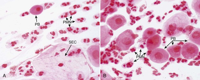
Although uncommon, there are other infectious causes of vaginitis. Three are briefly mentioned here because Gram stain of vaginal secretions may be helpful. First, Sobel described a number of premenopausal patients with a diffuse, exudative vaginitis with massive vaginal cell exfoliation, purulent vaginal discharge, and an occasional vaginal and cervical spotted rash. Laboratory findings included elevated pH of vaginal secretions. Also, numerous polymorphonuclear cells, an increased number of parabasal cells, the absence of gram-positive bacilli, and their replacement by occasional gram-positive cocci are observed on direct Gram stain (Figure 74-3). Basal cells appear as a result of the extensive exfoliation of epithelial cells. This clinical syndrome is referred to as desquamate inflammatory vaginitis. Symptoms associated with another disorder, lactobacillosis, resemble those of candidiasis and often follows antifungal therapy. Gram stain or wet mount typically reveals a large number of very long lactobacilli. These predominately anaerobic lactobacilli are 40 to 75 µ in length and are significantly longer than the average normal flora lactobacillus (5 to 15 µ). Finally, preexisting lesions due to other diseases may become secondarily infected with a mixed anaerobic flora of fusobacteria and spirochetes. This is referred to as fusiform-spirochete disease; this infection can progress rapidly. Gram stain examination reveals inflammatory cells in conjunction with gram-negative, fusiform bacterial morphotypes and spirochetes.


Cervicitis.

Polymorphonuclear neutrophils (PMNs) are normally present in the endocervix; however, an abnormally increased number of PMNs may be associated with cervicitis (inflammation of the cervix). Therefore, a purulent discharge from the endocervix can be observed in some cases of cervicitis. The endocervix is the site from which N. gonorrhoeae is most frequently isolated in women with gonococcal infections. In patients presenting with cervicitis, C. trachomatis can also be isolated; chlamydia have not been associated with vaginitis. Frequently, patients are infected with both pathogens. Because most women with cervicitis caused by gonococci or chlamydia are asymptomatic and cervical abnormalities are either subtle or absent in these women, an appropriate laboratory diagnosis to detect these organisms must be performed.


HSV and human papillomavirus (HPV) can also infect the cervix. In women with herpes cervicitis, the cervix is friable (bleeds easily) and may have ulcers. Affected patients may also have lower abdominal pain.


Anorectal Lesions.

As previously mentioned, because of the homosexual practice and increasingly common heterosexual practice of anal-genital intercourse, sites of infection in addition to those in the genital tract must be considered. The anorectum and pharynx are commonly infected with the classic STDs, including anal warts caused by HPV, as well as other viruses and parasites. Patients with symptoms of proctitis (inflammation of the rectum) caused by N. gonorrhoeae or C. trachomatis complain of itching, mucopurulent anal discharge, anal pain, bleeding, and tenesmus (painful straining during a bowel movement). Anorectal infection caused by HSV is associated with severe anal pain, rectal discharge, tenesmus, and systemic signs and symptoms such as fever, chills, and headaches.


In HIV-infected individuals and other immunocompromised patients, these infections tend to last longer, be more severe, and are more difficult to treat compared with infection in immunocompetent individuals. Anorectal lesions are common in HIV-infected patients and include anal condylomata, anal abscesses, and ulcers. Anal abscesses and ulcers can be due to various organisms, including CMV, Mycobacterium avium complex, HSV, Campylobacter spp., and Shigella, as well as traditional etiologic agents of STDs.

Stay updated, free articles. Join our Telegram channel

Aug 25, 2016 | Posted by in MICROBIOLOGY | Comments Off on Genital Tract Infections

Full access? Get Clinical Tree

Get Clinical Tree app for offline access