Genetics


Genetics


Genetics is the study of heredity—the passing of physical, biochemical, and physiologic traits from biological parents to their children. In this transmission, disorders can be transmitted and mistakes or mutations can result in disability or death.

The instructions for traits are carried within our genes. A gene is a segment of deoxyribonucleic acid (DNA) and serves as the template for eventual production of a protein. DNA is a double helix polymer (macromolecule) made up of individual units called nucleotides. Each nucleotide is composed of one sugar (deoxyribose), one phosphate, and one nitrogen-containing base. It’s estimated that there are over 3 billion nucleotides within our human genome. Each gene can contain hundreds to thousands of nucleotides; yet, genes make up less than 5% of our DNA. Each DNA double helix is tightly wound around histone proteins to form a chromosome. Every normal human cell (except reproductive cells) has 46 chromosomes, 22 paired chromosomes called autosomes, and 2 sex chromosomes (a pair of Xs in females and an X and a Y in males). A representation of a person’s individual set of chromosomes is called his karyotype. (See Normal human karyotype.)

Since 1990, the human genome has been studied to determine the exact order (sequence) of nucleotides in gene rich areas of the DNA. Human genome research continues to be of high importance in all areas of biomedical research as genes within the DNA sequence are identified, their function determined, and the consequences of genetic alterations and relationship to disease are studied. These areas of research continue to result in improved diagnostics and health outcomes for persons with or at risk for developing genetic diseases. (See The genome at a glance, page 94.)

A word of warning at the outset: For a wide variety of reasons, not every gene that might be expressed actually is expressed. Thus, the following chapter may seem to contain a great many “hedge” words, such as “may,” “perhaps,” and “some.” Genetic principles are based on studies of thousands of individuals. Those studies have led to generalities that are usually true, but exceptions occur. Genetics is a young science.


Genetic components

The DNA within our human genome consists of over 3 billion nucleotides. Each nucleotide contains one of four possible nitrogenous bases: adenine (A), thymine (T), guanine (G) or cytosine (C). The two strands of a DNA helix in a chromosome are joined at the bases by weak hydrogen bonds. Adenine joins with thymine and guanine joins with cytosine. The looseness of the bonds allows the strands to separate easily during cell division. (See DNA duplication: Two double helices from one, page 95.) The genes carry a code for each trait a person inherits, from blood type to eye color to body shape and myriad of other traits.

DNA ultimately controls the formation of essential substances throughout the life of every cell in the body, and it does this through the genes. A gene ultimately determines the linear sequence of an amino acid chain. The amino acid chain is modified to produce a specific
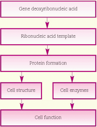
protein, which is necessary for cellular structure or function and contributes to a particular inherited trait, such as eye color or blood type. In the cell, a gene is transcribed within the nucleus into messenger ribonucleic acid (mRNA). This message leaves the nucleus and many different chemicals and cellular components within the cell cytoplasm are used to translate mRNA into an amino acid chain. A specific cellular component, called a ribosome, reads the message of three mRNA bases at a time. A sequence of three mRNA bases is called a codon, and there are 64 different codons within our genetic code. The ribosome reads the mRNA codons to determine and assemble the sequence of amino acids that will undergo further modification within the cell to become a functional protein. (See How genes control cell function, page 96.)


Trait transmission

Germ cells, or gametes (ovum and sperm), are one of two classes of cells in the body; each germ cell contains 23 chromosomes (called the haploid number) in its nucleus. All the other cells in the body are somatic cells, which are diploid, meaning they contain 23 pairs of chromosomes.

When ovum and sperm unite, the corresponding chromosomes pair up so that the fertilized cell and every somatic cell of the new person has 23 pairs of chromosomes in its nucleus.


GERM CELLS

The body produces germ cells through a type of cell division called meiosis. Meiosis occurs only when the body is creating haploid germ cells from their diploid precursors. Each of the 23 pairs of chromosomes in the germ cell separates so that, when the cell then divides, each new cell (ovum or sperm) contains one set of 23 chromosomes.

Most of the genes on one chromosome are identical or almost identical to the gene on its mate. (As we discuss later, each chromosome may carry a different version of the same gene.) The location (or locus) of a gene on a chromosome is specific and doesn’t vary from person to person.


Determining sex

Only one pair of chromosomes in each cell—pair 23—is involved in determining a person’s sex. These are the sex chromosomes; the other 22 numbered chromosome pairs are called autosomes. Females have two X chromosomes and males have one X and one Y chromosome.

Each gamete produced by a male contains either an X or a Y chromosome. When a sperm with an X chromosome fertilizes an ovum, the offspring is female (two X chromosomes); when a sperm with a Y chromosome fertilizes an ovum, the offspring is male (one X and one Y chromosome). Very rare errors in cell division can result in a germ cell that has no sex chromosome or two sex chromosomes. After fertilization with a gamete that contains a missing or extra sex chromosome, the zygote may have an XO, XXY, XXX, or XYY karyotype and still survive. Most other errors in sex chromosome division are incompatible with life.


MITOSIS

The fertilized ovum—now called a zygote—undergoes a type of cell division called mitosis. Before a cell divides, its chromosomes duplicate. During this process, the double helix of DNA separates into two chains; each chain serves as a template for constructing a new chain. Individual DNA nucleotides are linked into new strands with bases complementary to those in the originals. In this way, two identical double helices are formed, each containing one of the original strands and a newly formed complementary strand. These double helices are duplicates of the original DNA chain.

The cell cycle within somatic cells consists of alternations between interphase and mitosis —protein synthesis and DNA replication occur during interphase. Mitosis consists of four phases: prophase, metaphase, anaphase and telophase. Following telophase, cytokinesis occurs resulting in two new daughter cells, each genetically identical to the original and to each other. (See Five phases of mitosis, page 97.) Each of the two resulting cells likewise divides, and so on, eventually forming a many-celled human embryo. Thus, each cell in a person’s body (except ovum or sperm) contains an identical set of 46 chromosomes that are unique to that person.


Trait predominance

Each parent contributes one set of chromosomes (and therefore one set of genes) so that every offspring has two genes for every locus (location on the chromosome) on the autosomal chromosomes.

Some characteristics, or traits, are determined by one gene that may have many variants (alleles), such as the ability to roll the tongue. A person who has identical alleles on each chromosome is homozygous for that gene; if the alleles are different, they’re said to be heterozygous.

Others, called polygenic traits, require the interaction of one or more genes. Recent research has revealed that eye color is a polygenic trait. Three different genes at three different chromosome locations influence eye color. In addition, environmental factors may affect how a gene or genes are expressed.


AUTOSOMAL INHERITANCE

For unknown reasons, on autosomal chromosomes, one allele may be more influential than the other in determining a specific trait. The more powerful, or dominant, allele is more likely to be expressed in the offspring than the less influential, or recessive, allele. Offspring will express a dominant allele when one or both chromosomes in a pair carry it. A recessive allele won’t be expressed unless both chromosomes carry recessive alleles. For example, the
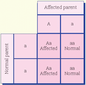
condition in which a person reflexively sneezes when quickly transitioning from a dark environment to intense bright light is thought to be dominant. A person who doesn’t experience this has two recessive alleles, whereas a person who does reflexively sneeze has either one or both dominant alleles for the trait.


SEX-LINKED INHERITANCE

The X and Y chromosomes aren’t literally a pair because the X chromosome is much larger than the Y. The male literally has less genetic material than the female, which means he has only one copy of most genes on the X chromosome. Inheritance of those genes is called X-linked. A man will transmit one copy of each X-linked gene to his daughters and none to his sons. A woman will transmit one copy to each child, male or female.

Inheritance of genes on the X chromosomes is different in another way. Some recessive genes on the X chromosomes act like dominants in males. Remember, males have an X and a Y chromosome in each somatic cell and few genes are shared between these two very different sex chromosomes. Therefore, genes on the X chromosome that don’t have partner genes on the Y chromosome will be expressed in males. In females, one of the two X chromosomes is randomly and permanently inactivated
in somatic cells during early embryogenesis. This phenomenon is called X-inactivation or Lyonization. X-inactivation ensures that females, like males, have one functional copy of the X chromosome in each body cell. The process of inactivating either the paternally contributed X or the maternally contributed X is random in each cell. Due to X-inactivation, one recessive allele will be expressed in some somatic cells and the partner allele (whether dominant or recessive) in other somatic cells. The most common example occurs not in people but in cats. Only female cats have calico (tricolor) coat patterns. Dark and orange hair color in the cat is carried on the X chromosome. Some hair cells in females express the brown allele and others, the orange color. The white color results from an autosomal gene.


MULTIFACTORIAL INHERITANCE

Some traits require a combination of two or more genes and environmental factors to be expressed. This is called multifactorial inheritance. Height is a classic example of a multifactorial trait. In general, the height of offspring will be in a range between the height of the two parents. But the combination of multiple genes contributed by each parent, nutritional patterns, health care, and other environmental factors also influence development. The better-nourished, healthier children of two short parents may be taller than either. Common health problems, such as obesity, diabetes, and hypertension, are associated with effects from multiple genes that are modified from environmental and lifestyle factors.


Pathophysiologic changes

Autosomal, sex-linked, and multifactorial disorders originate from damage to genes or chromosomes. Some defects arise spontaneously, whereas others may be caused by environmental teratogens.


GENE ERRORS

A rare change in genetic material is a mutation, which occurs in less than 1% of the population. A change that occurs with greater frequency is known as polymorphism. A mutation may occur spontaneously or after exposure of a cell to radiation, certain chemicals, or viruses. Mutations can occur anywhere in the genome—the person’s entire inventory of genes.

Every cell has built-in defenses against genetic damage. However, if a mutation isn’t identified or repaired, the mutation may produce a trait different from the original trait and is transmitted to offspring during reproduction. The mutation initially causes the cell to produce some abnormal protein that makes the cell different from its ancestors. Mutations may have no effect, they may change expression of a trait, and others change the way a cell functions. Some mutations cause serious or deadly defects, such as cancer or congenital anomalies.


Autosomal disorders

In single-gene disorders, an error occurs at a single gene site on the DNA strand. A mistake may occur in the copying and transcribing of a single codon (nucleotide triplet) through additions, deletions, excessive repetitions, or base changes.

Single-gene disorders are inherited in clearly identifiable patterns that are the same as those seen in inheritance of normal traits. Because every person has 22 pairs of autosomes and only 1 pair of sex chromosomes, most hereditary disorders are caused by autosomal mutations.


Autosomal dominant transmission usually affects male and female offspring equally. If one parent is affected, each child has one chance in two of being affected. If both parents are affected and each carries one dominant allele and one recessive allele for the disorder, they have affected or unaffected children. An example of this type of inheritance occurs in Marfan syndrome. (See Autosomal dominant inheritance.)

Autosomal recessive inheritance also usually affects male and female offspring equally. If both parents are affected, all of their offspring will be affected. If both parents are unaffected but are heterozygous for the trait (carriers of the defective gene), each child has one chance in four of being affected. If only one parent is affected, and the other is not a carrier, none of their offspring will be affected, but all will carry the defective gene. If one parent is affected and the other is a carrier, their offspring will have a 50% chance of being affected. (See Autosomal recessive inheritance.) Autosomal recessive disorders may occur when there is no family history of the disease.


Sex-linked disorders

Genetic disorders caused by genes located on the sex chromosomes are termed sex-linked disorders. Most sex-linked disorders are passed on the X chromosome, usually as recessive traits. Because males have only one X chromosome, a single X-linked recessive gene can cause disease to be exhibited in a male. Females receive two X chromosomes, so they can be homozygous for a disease allele, homozygous for a normal allele, or heterozygous (a carrier).

Most people who express X-linked recessive traits are males with unaffected parents. In rare
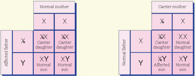
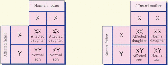
cases, the father is affected and the mother is a carrier. All daughters of an affected male will be carriers. Sons of an affected male will be unaffected, and the unaffected sons aren’t carriers. Unaffected male children of a female carrier don’t transmit the disorder. Hemophilia is an example of an X-linked inheritance disorder. (See X-linked recessive inheritance.)


Characteristics of X-linked dominant inheritance include evidence of the inherited trait in the family history. A person with the abnormal trait usually has one affected parent (except in the case of a new mutation occurring in a germ cell that conceived the individual). If the father has an X-linked dominant disorder, all his daughters and none of his sons will be affected. If a mother has an X-linked dominant disorder, each of her children has a 50% chance of being affected. (See X-linked dominant inheritance, page 100.)


Multifactorial disorders

Most multifactorial disorders result from the effects of several different genes and an environmental component. In polygenic inheritance, each gene has a small additive effect, and the effect of a combination of genetic errors in a person is unpredictable. Multifactorial disorders can result from a less-than-optimum expression of many different genes, not from a specific error.

Some multifactorial disorders are apparent at birth, such as cleft lip, cleft palate, congenital heart disease, anencephaly, clubfoot, and myelomeningocele. Others don’t become apparent until later, such as type 2 diabetes mellitus, hypertension, hyperlipidemia, most autoimmune diseases, and many cancers. Multifactorial disorders that develop during adulthood are often believed to be strongly related to environmental factors, not only in incidence but also in the degree of expression.


Environmental teratogens

Teratogens are environmental agents that can harm the developing fetus by causing congenital structural or functional defects. Teratogens may also cause spontaneous miscarriage, complications during labor and delivery, hidden defects in later development (such as cognitive or behavioral problems), or neoplastic transformations. (See Teratogens and associated disorders, page 101.)

Environmental factors of maternal or paternal origin include the use of chemicals (such as drugs, alcohol, or hormones), exposure to radiation, general health, and age. Maternal factors include infections during pregnancy, existing diseases, nutritional factors, exposure to high altitude, maternal-fetal blood incompatibility, and poor prenatal care.

The embryonic period—the first 8 weeks after fertilization—is a vulnerable time when specific organ systems are actively differentiating. Exposure to teratogens usually kills the embryo. During the fetal period, organ systems are formed and continue to mature. Exposure during this time can cause intrauterine growth retardation, cognitive abnormalities, or structural defects.


CHROMOSOME DEFECTS

Aberrations in chromosome structure or number cause a class of disorders called congenital anomalies, or birth defects. The aberration may be loss, addition, or rearrangement of genetic material. If the remaining genetic material is sufficient to maintain life, an endless variety of clinical manifestations may occur. Most clinically significant chromosome aberrations arise during meiosis. Meiosis is an incredibly complex process that can go wrong in many ways. Potential contributing factors include maternal age, radiation, and use of some therapeutic or recreational drugs.

Translocation, the shifting or moving of chromosomal material, occurs when chromosomes split apart and rejoin in an abnormal arrangement. The cells still have a normal amount of genetic material, so often there are no visible abnormalities. However, the children of parents with translocated chromosomes may have serious genetic defects, such as monosomies or trisomies. Parental age doesn’t seem to be a factor in translocation.


Errors in chromosome number

During both meiosis and mitosis, chromosomes normally separate in a process called disjunction. Failure to separate, called nondisjunction, causes an unequal distribution of chromosomes between the two resulting cells. If nondisjunction occurs during mitosis soon after fertilization, it may affect all the resulting cells. Gain or loss of chromosomes is usually caused by nondisjunction of autosomes or sex chromosomes during meiosis. The incidence of nondisjunction increases with maternal age. (See Chromosomal disjunction and nondisjunction, page 103.)

The presence of one chromosome less than the normal number is called monosomy; an autosomal monosomy is nonviable. The presence of an extra chromosome is called a trisomy, the most common of which are trisomies 21, 18, and 13. Children with trisomy 18 or 13 usually don’t survive beyond the first months of life. A mixture of both trisomic and normal cells results in mosaicism, which is the presence of two or more cell lines in the same person. The effect of mosaicism depends on the proportion and anatomic location of abnormal cells.


Disorders

This section discusses disorders in the context of their pattern of inheritance as well as environmental factors. The alphabetically listed disorders have these patterns of inheritance:

♦ Autosomal recessive—cystic fibrosis, sickle cell anemia, Tay-Sachs disease

♦ Autosomal dominant—Marfan syndrome

♦ X-linked recessive—hemophilia, fragile X syndrome

♦ Polygenic multifactorial—cleft lip and cleft palate, neural tube defects

♦ Chromosome number—Down syndrome, Klinefelter syndrome.


CLEFT LIP AND CLEFT PALATE

Cleft lip and cleft palate may occur separately or in combination. They originate in the second month of pregnancy if the front and sides of the face and the palatine shelves fuse imperfectly. Cleft lip with or without cleft palate occurs twice as often in males than in females. Cleft palate without cleft lip is more common in females.

Cleft lip deformities can occur unilaterally, bilaterally or, rarely, in the midline. Only the lip may be involved, or the defect may extend into the upper jaw or nasal cavity. (See Types of cleft deformities, page 104.)

Incidence is highest in children with a family history of cleft defects.

Cleft lip with or without cleft palate occurs in about 1 in 1,000 births among Whites; the incidence is higher in Asians (1.7 in 1,000) and Native Americans (more than 3.6 in 1,000) but lower in Blacks (1 in 2,500).


Causes

♦ Chromosomal or Mendelian syndrome (cleft defects are associated with more than 300 syndromes)

♦ Exposure to teratogens during fetal development

♦ Combined genetic and environmental factors (accounts for 75% of isolated cleft cases)


Pathophysiology

During the second month of pregnancy, the front and sides of the face and the palatine shelves develop. Because of a chromosomal abnormality, exposure to teratogens, genetic abnormality, or environmental factors, the lip or palate fuses imperfectly.

The deformity may range from a simple notch to a complete cleft. A cleft palate may be partial or complete. A complete cleft includes the soft palate, the bones of the maxilla, and the alveolus on one or both sides of the premaxilla.

A double cleft is the most severe of the deformities. The cleft runs from the soft palate forward to either side of the nose. A double cleft separates the maxilla and premaxilla into freely moving segments. The tongue and other muscles can displace the segments, enlarging the cleft.


imageIsolated cleft palate is more commonly associated with other congenital defects than isolated cleft lip with or without cleft palate. The constellation of U-shaped cleft palate, mandibular hypoplasia, and glossoptosis is known as Pierre Robin syndrome, or Robin syndrome. It can occur as an isolated defect or one feature of many different syndromes; therefore, a comprehensive genetic evaluation is suggested for infants with Robin sequence. Because of the mandibular hypoplasia and glossoptosis, careful evaluation and management of the airway are mandatory for infants with Robin sequence.


Signs and symptoms

♦ Obvious cleft lip or cleft palate due to incomplete fusion of the lip or palate

♦ Feeding difficulties due to incomplete fusion of the palate


Complications

♦ Malnutrition, because the abnormal palate affects nutritional intake

♦ Hearing impairment, often due to middle-ear damage or recurrent infections

♦ Permanent speech impediment, even after surgical repair


Diagnosis

♦ Clinical presentation, obvious at birth

♦ Prenatal targeted ultrasound


Treatment

♦ Surgical correction of cleft lip in the first few days of life to permit sucking, or delayed for 8 to 10 weeks (sometimes as long as 6 to 8 months) to allow the infant to grow and mature, thereby minimizing surgical and anesthesia risks, ruling out associated congenital anomalies, and allowing time for parental bonding

♦ Orthodontic prosthesis to improve sucking

♦ Surgical correction of cleft palate at 12 to 18 months, after the infant gains weight and is infection-free

♦ Speech therapy to correct speech patterns

♦ Use of a contoured speech bulb attached to the posterior of a denture to occlude the nasopharynx when a wide horseshoe defect makes surgery impossible (to help the child develop intelligible speech)

♦ Adequate nutrition for normal growth and development

♦ Use of a large soft nipple with large holes, such as a lamb’s nipple, to improve feeding patterns and promote nutrition

image
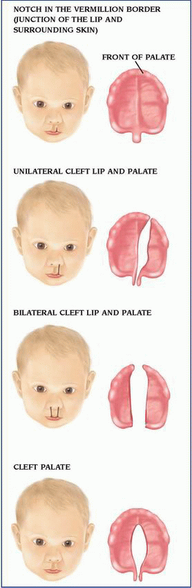
Daily use of folic acid before conception and during pregnancy decreases the risk for isolated (not associated with another genetic or congenital malformation) cleft lip or palate by up to 25%. Women of childbearing age should be encouraged to take a daily multivitamin containing folic acid until menopause or until they’re no longer fertile.

Aug 27, 2016 | Posted by in PATHOLOGY & LABORATORY MEDICINE | Comments Off on Genetics

Full access? Get Clinical Tree

Get Clinical Tree app for offline access