Fig. 17.1
Adult with multiple FDE due to a Cephalosporin
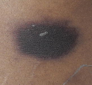
Fig. 17.2
Recurrent localized FDE. Hyperpigmented patch with surrounding violaceous erythema
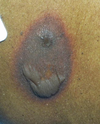
Fig. 17.3
Bulla formation in localized FDE
The first time an individual is exposed to a drug, the FDE may not appear for up to 1 week. With subsequent exposure to the same drug, the patches and plaques can develop as early as 30 min following drug exposure, with most reactions appearing within 8–24 h. Patients do not normally report constitutional symptoms; however, pruritus and pain at the site of the FDE are two commonly reported symptoms that can precede the appearance of the lesions.
FDEs have a predilection for areas with thin skin, such as the lips, genitalia, and perianal regions, although they have also been reported on the trunk, palms, soles, and web spaces of the hands and feet (Figs. 17.4 and 17.5). Mucosal involvement often leads to isolated erosions overlying the erythematous macules and patches. Despite these commonly reported sites of FDEs, lesions can appear on any area of the skin, mucosa, or mucocutaneous junction. Case reports have described a Koebner-like phenomenon in patients with FDEs, with lesions occurring over areas of previous trauma.
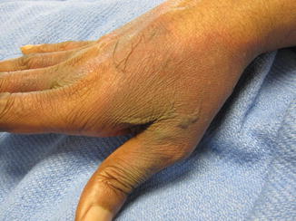

Figs. 17.4 and 17.5
FDE involving the bilateral hands
FDEs occur with equal prevalence among males and females, and cases have been documented in all age groups. In adults, morbilliform rashes make up around 95 % of cutaneous drug reactions, while in children FDEs account for up to 25 % of cutaneous drug reactions. The patient history will normally reveal recent introduction of a new analgesic, antibiotic, sedative, or anticonvulsant. These cases are easier to identify in the hospital setting due to better documentation of the patient’s medication regimen. In outpatient clinics, patients may not be seen until the second or third FDE occurrence, especially if the lesion is not symptomatic, which makes identification of the causative drug difficult. With the introduction of new analgesics to the market, there is an increasing report of “polysensitivity.” For example, a patient may report their first FDE with one analgesic and subsequently avoid that analgesic. However, upon exposure to a second analgesic, they have an FDE recurrence despite the use of a different medication. The cross-reactivity in this class of drugs has not been fully explored, although it is likely that the medications have similar ingredients to which the patient is sensitive.
Special Variants: Localized FDE (Non-pigmenting)
The non-pigmenting fixed drug eruption (NPFDE) is a variant of FDE that is more common in pediatric patients. NPFDEs evolve over the first 24 h with similar clinical and histological characteristics of FDE. The non-pigmenting term refers to the recovery phase of these lesions. Although NPFDEs will recur at the same site as the initial presentation like an FDE, the non-pigmenting variety does not leave a well demarcated patch of hyperpigmentation characteristic of classic FDE. The original well-circumscribed patch or plaque slowly fades over 2–3 weeks. Very few cases have been described, and the mechanism by which complete resolution of the lesions between exposures occurs is not well known. In addition, while FDEs are almost always asymmetrical, occurring in an unpredictable pattern on almost any exposed skin or mucosa, NPFDEs have been described as symmetrical, well-demarcated, erythematous patches and plaques.
Fixed drug eruptions in children are not as well described in the literature in comparison to the adult population. It has been suggested that FDEs occur more frequently in the pediatric population and may possibly be one of the most common drug eruptions in children, however, often the lesions go misdiagnosed. Pediatricians commonly encounter urticarial eruptions and morbilliform rashes following ingestion of certain medications, but the well-demarcated lesions in a FDE can be missed if it is attributed to other causes after a single exposure to the offending agent, since recurrence at the same site is the hallmark of FDE. Acetaminophen and sulfonamides are the most cited drugs causing FDE in children, while pseudoephedrine is the most common inciting agent in adults. Unlike adults, tetracycline is not a well-known cause of FDE in children, likely due to the fact that this antibiotic is not regularly administered to children under 8 years of age because of its side effects.
Diagnosis and treatment in children is similar to adults. Patch testing and oral re-challenge can confirm the offending drug, and symptomatic treatment can control pain and itching, but do not expedite the healing process.
Generalized Bullous FDE
In severe generalized bullous FDE (GBFDE), the patches and plaques can cover large surface areas, mimicking toxic epidermal necrolysis and Stevens-Johnson Syndome (TEN/SJS) with development of central flaccid bullae or vesicles within the edematous plaques. GBFDE appears on both the trunk and extremities, and the patches and plaques remain well-demarcated, as in other FDE variants (Fig. 17.6). Like TEN/SJS, GBFDE may present with constitutional symptoms, although not to the same degree as the former. GBFDE is also less likely to involve mucosal surfaces and will usually resolve within 7–14 days once the drug has been discontinued. Nonetheless, the presentation can be quite severe, and a recent review noted that mortality rates may approach that of TEN/SJS.
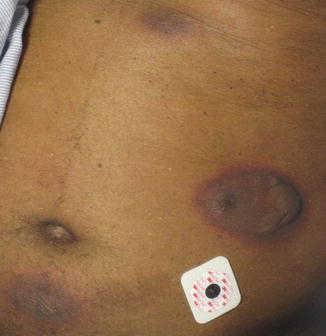
Fig. 17.6
Multiple bullous FDE due to sulfamethoxazole/trimethoprim
Inverse FDE
Fixed drug eruptions with an inverse or flexural distribution have been reported and are thought to be variants of the symmetrical drug-related intertriginous and flexural exanthema (SDRIFE). The literature is limited regarding this presentation, but Ozkaya and Babuna described a case of a 12-year-old boy who presented with an amoxicillin-induced drug eruption who developed large, well-demarcated, erythematous symmetrical plaques in his gluteal cleft and flexor regions. The lesions resolved with FDE-characteristic hyperpigmentation, and the plaques reappeared with oral re-challenge of amoxicillin at a sub-therapeutic dose. The symmetry of his lesions in predominantly flexor areas, as well as the gluteal cleft involvement, was more consistent with SDRIFE; however, the well-demarcated lesions, residual hyperpigmentation, and site-specific recurrence were more consistent with FDE. It is likely that overlap between both eruptions exists, as illustrated in Figs. 17.7 and 17.8.
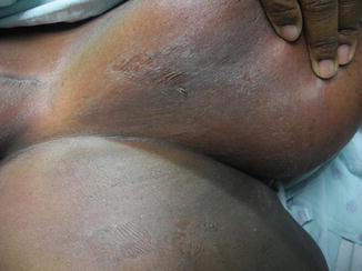
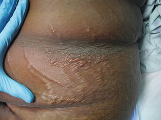
Figs. 17.7 and 17.8
Involvement of flexural areas and skin folds in an inverse FDE or SDRIFE-FDE overlap
Oral–Genital Fixed Drug Eruptions
Oral–genital mucosal FDEs can occur as solitary lesions or in addition to lesions affecting other areas of skin. The two main offending agents are naproxen and trimethoprim-sulfamethoxazole. The latter drug has a predilection for lesions on the dorsal surface of the tongue, while the former affects the dorsum of the tongue and the hard palate with equal prevalence (Fig. 17.9). However, lesions have been reported on the mucosal lip, buccal mucosa, and gingiva as well.
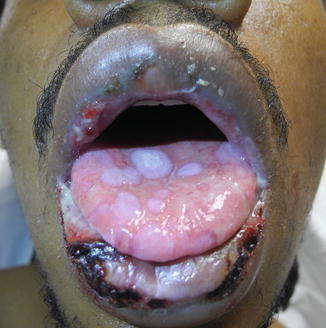
Fig. 17.9
Oral FDE due to Ibuprofen, involving the dorsal tongue
FDE involving the genital mucosa is well described, and commonly presents as an itchy or painful erythematous plaque that may become bullous or eroded. The most commonly involved site in male patients is the glans penis; however, the shaft and scrotum may also be affected (Fig. 17.10). Previous reviews have suggested that and trimethoprim-sulfamethoxazole and tetracycline are the most common causal agents.

Fig. 17.10
FDE due to sulfamethoxazole/trimethoprim, involving the penile shaft
Multiple case series of genital FDE have included only a small number of female patients, suggesting that the entity is likely underreported in women. Female patients with genital FDE are likely to present to their primary care physician or gynecologist and are often diagnosed with another condition. Additionally, several cases of genital FDE have been reported in patients who were not ingesting the drug, but whose sexual partner was taking the offending drug, with the patient being exposed to the drug through sexual contact. In a recent series of patients with vulvar FDE, the most common clinical presentation was bilaterally symmetrical erythematous vulvitis, involving the labia minora and majora and extending to the perineum (Fig. 17.11). Involvement of the inner thighs and perianal area can also occur. Chronic erosive mucositis is another presentation of genital FDE. The most common associated drugs are NSAIDs (ibuprofen, cyclo-oxygenase-2 inhibitors and 3-hydroxy-3-methylglutaryl-CoA reductase inhibitors). Because NSAIDs are one of the most common offenders, female patients may present with a cyclical pattern of oral-mucosal lesions associated with treating dysmenorrhea. The condition is often misdiagnosed as other cyclical diseases, such as herpes simplex virus infection or Behcet’s disease.
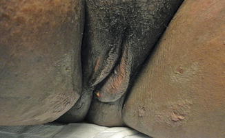
Fig. 17.11
FDE involving the labia majora, extending down the bilateral inner thighs and perineal area
Diagnosis is difficult when lesions are not associated with classical FDE lesions on the skin because they do not leave the classic residual hyperpigmentation following their resolution. The lesions also vary in appearance, including bullous/erosive and aphthous forms or superficial erosion with an erythematous base. The bullous/erosive pattern is most frequent, especially on the aforementioned preferred mucosal locations. Patients report pain and burning as the main symptoms, which resolve with discontinuation of the offending drug.
Diagnosis
Patients should be questioned regarding all drugs they have ingested, including over-the-counter and prescribed medications. Nutritional supplements, herbal products, and diet aids are possibilities as well. Patients tend to overlook medications that they have been taking sporadically over years. Often asking a patient whether they take any medications for specific conditions or symptoms such as headache, muscle pains, constipation, urinary tract infections, and menstrual cramps may yield a more accurate history. Biopsy can be helpful in atypical cases such as in generalized or bullous FDE, for those with oral or genital involvement, and those in whom the diagnosis is unclear. The presence of systemic symptoms such as fever, arthralgia, or malaise may also be indications for biopsy.
Stay updated, free articles. Join our Telegram channel

Full access? Get Clinical Tree


