Fibromatosis
Key Facts
Terminology
Neoplastic proliferation of fibroblasts that is locally infiltrative but which does not metastasize
Etiology/Pathogenesis
Some cases are associated with familial adenomatous polyposis (including Gardner and Turcot syndromes) and germline mutations in APC gene
Sporadic cases are also associated with mutations in APC gene (20% of tumors) or β-catenin gene (50% of tumors)
Minority of cases are associated with prior trauma or surgery
Clinical Issues
Rare; only 0.2% of breast tumors
Most patients present with palpable mass or radiologic findings that closely mimic invasive carcinoma
Local recurrence is common within 1st 3 years (20-30%), even with uninvolved margins
Microscopic Pathology
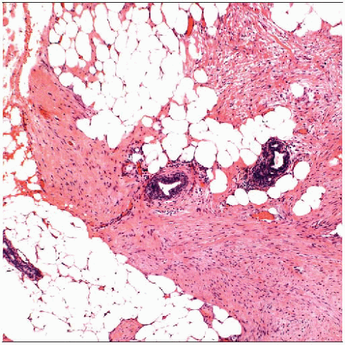
Long fascicles of spindle cells that infiltrate around normal ducts and lobules and may infiltrate into muscle
Ancillary Tests
Fibromatosis characteristically shows aberrant nuclear β-catenin positivity and absence of CD34
 Fibromatosis is characterized by a proliferation of bland spindle cells organized in long fascicles that surround normal ducts and lobules and infiltrate into adipose tissue and skeletal muscle. |
TERMINOLOGY
Synonyms
Mammary fibromatosis, aggressive fibromatosis
Desmoid tumor, desmoid-type fibromatosis, extraabdominal desmoid tumor
Definitions
Neoplastic proliferation of fibroblasts and myofibroblasts that is locally infiltrative but does not metastasize
ETIOLOGY/PATHOGENESIS
Genetic Causes
Germline mutations
Familial adenomatous polyposis (FAP; includes Gardner syndrome and Turcot syndrome) is due to germline mutation in adenomatous polyposis coli (APC) gene
APC gene regulates β-catenin
Fibromatosis may occur in breast in patients with FAP, but fibromatosis is more common at other body sites
Abnormal regulation of β-catenin can result in accumulation of protein in nucleus, in addition to normal location in cytoplasm
Fibromatosis at any site associated with FAP shows nuclear β-catenin positivity in 67% of cases
Somatic mutations
In sporadic cases, mutations in APC gene are present in 20% of tumors
Mutations in β-catenin gene in 50% of sporadic tumors
80% of sporadic cases of fibromatosis are positive for nuclear β-catenin
Other Causes
Trauma or prior surgery
In minority of cases, fibromatosis occurs in area of prior trauma or surgery (e.g., within capsule of breast implant)
CLINICAL ISSUES
Epidemiology
Incidence
Rare; only 0.2% of breast tumors
Age
Most patients are women in their 40s; however, any age can be affected
Presentation
Most patients present with palpable mass or radiologic findings that closely mimic invasive carcinoma
Tumor can be fixed to pectoralis muscle or retract skin
Almost 1/2 of patients have prior history of breast surgery
Masses are usually painless
Tumors are irregular in shape
Treatment
Tumor should be completely excised with an attempt at uninvolved margins
However, margin status does not accurately predict risk of local recurrence
Radiation has been used for some patients; however, effectiveness of this treatment is uncertain
Adjuvant treatment with cytotoxic therapy or hormonal therapy has been used
Too few cases reported to determine effectiveness of these treatments
Prognosis
Local recurrence is common within 1st 3 years (20-30%), even with uninvolved margins
Younger patients with larger tumors are at increased risk for local recurrence
Not all patients with positive margins recur; therefore, careful observation with resection at time of recurrence is possible approach
Histologic features do not identify tumors most likely to recur
IMAGE FINDINGS
Mammographic Findings
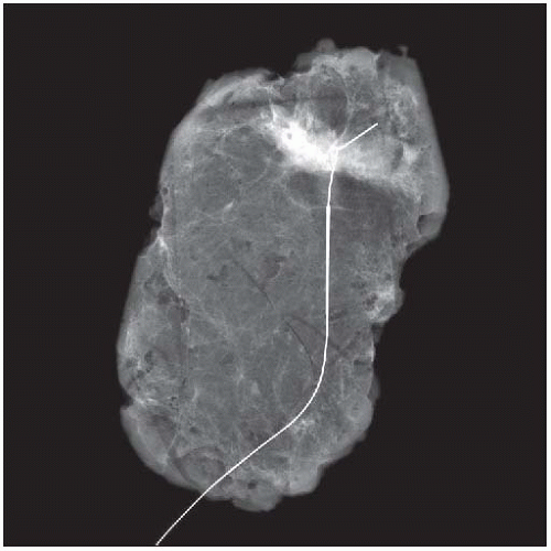
Irregular dense mass; calcifications are not typical
Most typical location is close to, or arising from, pectoralis muscle fascia
If lesion is very large, breast can appear retracted and reduced in size
Ultrasonographic Findings
Irregular mass with thick echogenic rim
MR Findings
Mass-forming lesion with irregular margins
Variable enhancement kinetics
Some reports of benign gradual enhancement pattern
Other enhancement patterns mimic malignancy
MACROSCOPIC FEATURES
General Features
Firm to rubbery white mass
Borders can appear circumscribed or irregular, or mass can be ill defined
Size can range from 1 cm to > 10 cm
MICROSCOPIC PATHOLOGY
Histologic Features
Long fascicles of spindle cells infiltrate around normal ducts and lobules and may infiltrate into muscle
Cells are bland with minimal nuclear pleomorphism
Mitoses are rare
Lymphocytic aggregates are typically at periphery of tumor
Stromal collagen bundles may be prominent
Spindle cells consist of fibroblasts and myofibroblasts
Diagnosis is often difficult to make on core needle biopsy
ANCILLARY TESTS
Immunohistochemistry
Majority of tumors have aberrant nuclear β-catenin positivity, as well as typical cytoplasmic positivity
Not specific for fibromatosis and may be seen in other benign and malignant tumors
Tumors frequently have subpopulation of cells positive for muscle markers: Desmin, actin, calponin
Cells are negative for CD34; only other breast stromal tumor negative for CD34 is nodular fasciitis
Spindle cell carcinoma should be excluded by use of broad spectrum keratins, including basal-like keratins
Cells are negative for estrogen and progesterone receptors
DIFFERENTIAL DIAGNOSIS
Spindle Cell Carcinoma (Including Fibromatosis-like Metaplastic Carcinoma)
Carcinomas generally have greater degree of cellular pleomorphism and more frequent mitoses
Fibromatosis-like metaplastic carcinomas can closely resemble fibromatosis
Carcinomas will be immunoreactive for cytokeratins; it may be necessary to use basal keratins 14 and 17
Stay updated, free articles. Join our Telegram channel

Full access? Get Clinical Tree


