Extramedullary Hematopoiesis


EMH With Megakaryocyte
This is a skin biopsy specimen with extramedullary hematopoiesis (EMH), as evidenced by the large megakaryocyte image.


image
EMH With Erythroid Cells
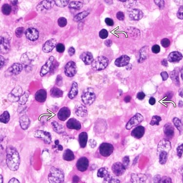
This cutaneous lesion contains small clusters of nucleated red blood cells image, which were positive for a hemoglobin A immunostain (not shown).

image
Factor VIII Immunohistochemistry
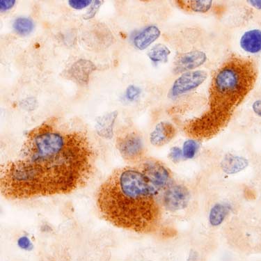
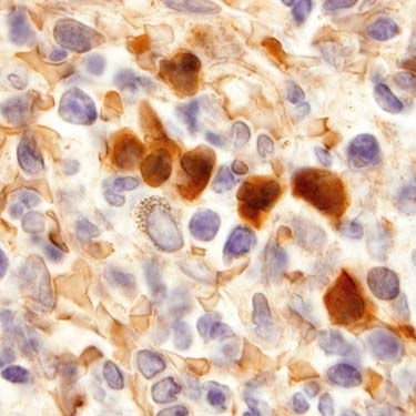
This is a factor VIII immunostain in EMH of the skin. The megakaryocytes are positive for factor VIII. Other megakaryocyte markers include CD61, CD34, and LMP.

image
Hemoglobin A Immunohistochemistry
This is a hemoglobin A immunostain in a skin biopsy specimen with EMH; the stain is highlighting erythroid cells.


TERMINOLOGY


Abbreviations


• Extramedullary hematopoiesis (EMH)


Definitions


• Formation of blood cells (including nucleated red blood cells, lymphocytes, and megakaryocytes) outside of bone marrow
image Occurs most commonly in liver and spleen

– Also found in other organs, including lymph nodes, kidney, adrenal, gastrointestinal tract, lung, breast, central nervous system

image Hematopoiesis occurs in skin early in embryonic life


ETIOLOGY/PATHOGENESIS


Viral Infection


• Has been reported in skin in neonates with intrauterine viral infections including
image Cytomegalovirus

image Coxsackievirus

image Rubella


Hematopoietic Disorders


• In neonates, EMH occurs in congenital hematologic disorders including
image Hemolytic disease of newborn

image Twin transfusion syndrome

• In adults, EMH is seen with hematopoietic disorders
image All myeloproliferative neoplasms are associated with EMH
– Primary myelofibrosis is most common, especially in patients who have had splenectomy

Only gold members can continue reading. Log In or Register to continue

Stay updated, free articles. Join our Telegram channel

Apr 24, 2017 | Posted by in PATHOLOGY & LABORATORY MEDICINE | Comments Off on Extramedullary Hematopoiesis

Full access? Get Clinical Tree

Get Clinical Tree app for offline access