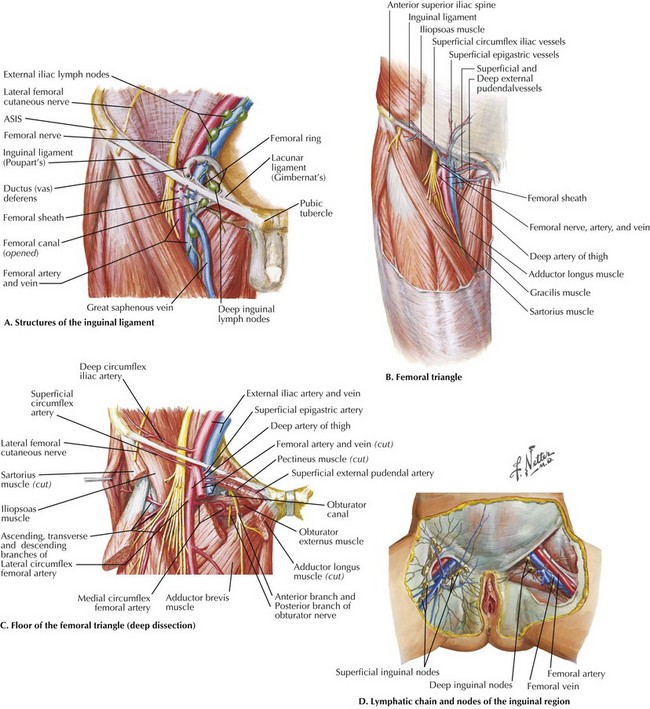
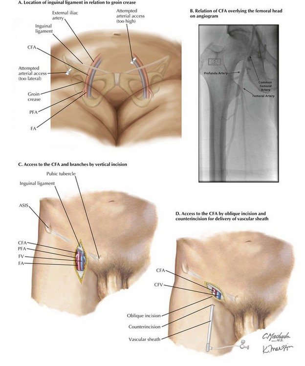
Chapter 37 The CFA measures approximately 5 cm and is the direct continuation of the external iliac artery, distal to the inguinal ligament. The femoral neurovascular bundle emerges from the retroperitoneum of the pelvis at the inguinal level on its way into the thigh. Just as the umbilicus is located toward the midline, the mnemonic NAVEL refers to these structures deep to the inguinal ligament, from lateral to medial: femoral Nerve, common femoral Artery (CFA), common femoral Vein (CFV), Empty space in femoral canal containing lymph vessels and lymph nodes, and lacunar Ligament (Fig. 37-1, A). The inguinal (Poupart’s) ligament, which spans from the anterior superior iliac spine (ASIS) to the pubic tubercle, also forms the superior border of the femoral triangle (Fig. 37-1, B). The lateral and medial borders are the sartorius and adductor longus muscles, respectively. The femoral triangle appears as a depressed plane on thigh flexion and external rotation and contains the femoral neurovascular bundle, including the two major CFA branches, the femoral and profunda femoris (deep femoral) arteries and their corresponding veins. Proximally, deep to the NAVEL structures, the floor of the triangle comprises, from lateral to medial, the iliopsoas and pectineus muscles and pectineal ligament condensation on the superior pubic ramus (Fig. 37-1, C). The roof of the triangle is formed by fascia lata of the thigh, which has an oval opening at the superomedial aspect (fossa ovalis) through which the greater saphenous vein and lymphatic vessels join the deeper structures. Lymphatic vessels and lymph nodes are oriented parallel to the CFA and CFV, as well as along the inguinal ligament (Fig. 37-1, D). The location of CFA for percutaneous access is usually guided by palpation of its pulse, typically located just medial and inferior to the midpoint of the inguinal ligament. A common misconception that leads to low arterial sticks and possible injury to the femoral or profunda artery is that the groin crease directly corresponds to the inguinal ligament (Fig. 37-2, A). The inguinal ligament is two to three fingerbreadths cephalad to the crease and is most reliably identified by bony landmarks (ASIS, pubic tubercle) on palpation or fluoroscopy. The CFA usually overlies the medial two thirds of the femoral head (Fig. 37-2, B). These guidelines are helpful when the CFA is pulseless (heavy calcification, severe stenosis) or indiscernible (body habitus, obesity).
Exposure of the Common Femoral Artery and Vein
Femoral Anatomy
Surgical Principles
![]()
Stay updated, free articles. Join our Telegram channel

Full access? Get Clinical Tree


Basicmedical Key
Fastest Basicmedical Insight Engine