Excisions
INTRODUCTION
Indications for Procedure
Breast excisions include all procedures with intent to remove entire lesion, but not entire breast
Excision is general term for any procedure that does not involve removal of entire breast
Biopsy: Used in similar fashion as excision but also includes special types of biopsies such as core needle biopsies or incisional biopsies
Lumpectomy: Procedure to remove a palpable mass; also used to generally refer to removal of nonpalpable lesions
Quadrantectomy: Removes an entire quadrant of breast
Partial mastectomy: Used synonymously with excisions
Incisional biopsies are less common procedures used to sample, but not completely remove, large lesions
In majority of cases, core needle biopsies are used in place of incisional biopsies
Excisions often result in better cosmetic results than mastectomy
Performed for initial diagnosis of lesions, treatment of symptoms, and treatment of carcinomas
Initial Investigation of a Lesion
Breast lesions suspicious for carcinoma require biopsy for evaluation
Many lesions can be evaluated using core needle biopsy
Some lesions are not amenable to needle biopsy
Location near nipple or deep in breast
Patient unable to remain immobile during procedure
Lesion difficult to visualize using digital mammography
Treatment of a Symptom
Some patients have symptoms that may be treated with excision
Palpable masses causing cosmetic changes or pain
Nipple discharge
Inflammatory lesions causing pain ± fistula tracts
If due to infectious organisms, incision and drainage may be necessary for cure
Breast-Conserving Therapy (BCT) for Cancer
Patients with cancer can generally be treated with BCT
Eligibility depends on size and location of cancer
Survival outcomes of BCT with radiation therapy are similar to those of mastectomy
Must be able to maintain acceptable cosmesis
Patients must be candidates for radiation therapy
Patient preference is also important consideration
Initial diagnosis of cancer by core needle biopsy aids in patient management
Subsequent excision is often larger and more likely to achieve negative margins
If clear margins cannot be achieved, mastectomy may be better option
For invasive carcinomas, lymph node sampling can be performed in same procedure
Standard of care is also to treat patients with radiation therapy to reduce risk of local recurrence
SPECIMEN PROCESSING
Requisition Form
In addition to information that should be provided with all breast specimens, the following information is necessary for gross examination of specimen
Type of lesion(s) targeted for removal
Palpable mass
Imaging finding (mammographic, ultrasound, or MR)
Duct excision for nipple discharge
Orientation of specimen
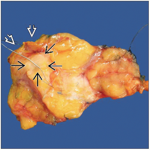
Designations of sutures or clips used to mark specific margins (all 6 margins should be identifiable)
If adequate information is not provided, additional information should be requested from surgeon
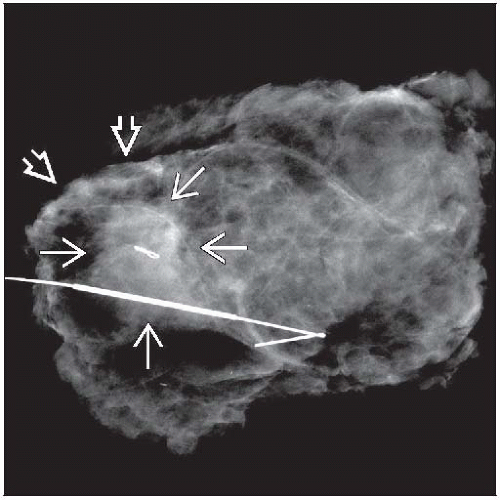
Specimen Radiograph
If specimen was excised for radiologic lesion, a specimen radiograph should be received with specimen
Radiologist’s interpretation should be available to pathologist
States whether or not lesion is present
Identifies type of lesion (e.g., mass or calcifications)
Often helpful for radiologist to circle or otherwise indicate location of lesion
Radiograph should be oriented with respect to specimen
4 margins can be identified on radiograph
Often provides useful information about closest margin to targeted lesion
Surgeons may use radiologic distance to margins to perform a reexcision if diagnosis of carcinoma has been made previously by core needle biopsy or fine needle aspiration
However, DCIS at margins is not usually evident by radiography
Radiograph can be annotated to designate margins and sites of tissue sampling
Likely location of targeted lesion in specimen can be approximated using shape of specimen and location of wire
Commercial specimen holders with grids are available; these help localize lesions in specimens
Stay updated, free articles. Join our Telegram channel

Full access? Get Clinical Tree


