Erythema Multiforme
DIAGNOSTIC SUMMARY
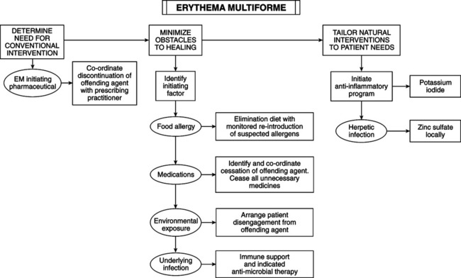
• Sudden onset of symmetric, erythematous, edematous, macular, papular, urticarial, bullous, or purpuric skin lesions.
• Evolves into “target lesions” (“bull’s eye” lesions with clear centers and concentric erythematous rings).
• Characteristic first site: dorsum of hand.
• Characteristic distribution: extensor surfaces of extremities with relative sparing of head and trunk.
• Rare oral manifestations: range from tender superficial erythematous and hyperkeratotic plaques to painful, deep, hemorrhagic bullae and erosions.
GENERAL CONSIDERATIONS
Erythema multiforme (EM) includes a wide range of expressions, from exclusive oral erosions (oral EM) to mucocutaneous lesions ranging from mild (EM minor) to severe multiple mucosal membranes (EM major, Stevens-Johnson syndrome [SJS]) or large area of total body surface (toxic epidermal necrolysis [TEN]). But the term EM is not accepted worldwide; various clinical categories have overlapping features. EM minor, EM major, SJS, and TEN differ in severity and expression. all variants share two common features: cutaneous target lesions and satellite cell or more widespread necrosis of epithelium. Suspected etiologic factors: herpes simplex or Mycoplasma are linked to 90% of cases of EM minor; SJS and TEN (80% of cases) arise from drugs: anticonvulsants, sulfonamides, nonsteroidal antiinflammatories, and antibiotics. Vaccines (e.g., Vaccinia, bacille Calmette-Guérin, polio), food allergy, and other infectious organisms have all induced EM minor. Common factor: highly reactive, polymorphonuclear leukocyte oxygen intermediates trigger hypersensitivity and immune system–mediated tissue damage.


Stay updated, free articles. Join our Telegram channel

Full access? Get Clinical Tree

