Epithelioid Sarcoma
Cyril Fisher, MD, DSc, FRCPath
Key Facts
Terminology
Malignant mesenchymal tumor resembling carcinoma or granuloma
Shows predominantly epithelial but also mesenchymal differentiation
Occurs in classical and proximal aggressive forms
Etiology/Pathogenesis
Some cases have abnormalities of chromosome 22q
Clinical Issues
Most common in distal extremities, especially hand and forearm
More frequent in males
Subcutaneous nonhealing ulcer or deep mass
High-grade sarcoma
Persistent and multiple recurrences
50% metastasize
Macroscopic Features
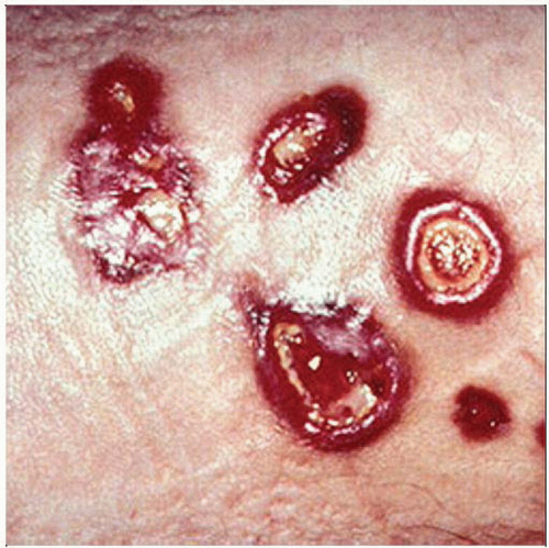
Raised “sealing-wax” margins around ulcer
Microscopic Pathology
Central necrosis
Transition to spindle cells at periphery
Nuclei bland or mildly atypical, resembles granuloma
Ancillary Tests
CK(+) and EMA(+)
CD34(+) in 50% of cases
S100 protein(-), desmin(-) and INI1(-)
Diagnostic Checklist
Granulomatous lesion: Look for CK positivity
 Clinical photograph shows several rounded lesions with raised red margins and central “targetoid” ulceration on the forearm of a 23-year-old man. Nonhealing ulcers are typical of epithelioid sarcoma. |
TERMINOLOGY
Abbreviations
Epithelioid sarcoma (ES)
Synonyms
Epithelioid cell sarcoma (no longer recommended)
Definitions
Malignant mesenchymal tumor resembling carcinoma or granuloma, which shows predominantly epithelial but also mesenchymal differentiation
ES occurs in classical and proximal (aggressive, large cell, or rhabdoid) forms
ETIOLOGY/PATHOGENESIS
Genetic Factors
Some cases have abnormalities of chromosome 22q
Rare association with neurofibromatosis type 2
CLINICAL ISSUES
Epidemiology
Incidence
Rare
Accounts for about 1% of all soft tissue sarcomas
Classic ES
Most common in distal extremities, especially hand and forearm
Head and neck
Penis, vulva
Proximal ES
Proximal limb girdle
Axial locations: Perineum, pelvis, mediastinum
Trunk: Chest wall
Age
Any age
Classic ES
Mostly 2nd-4th decades
Proximal ES
Median age 40 years (range 13-80 years)
Gender
More frequent in males
Presentation
Slow growing
Subcutaneous mass
Ulcer
Classic ES
Dermal or subcutaneous nodule
Nonhealing ulcer
Proximal ES
Subcutaneous or deep mass
Can appear more rapidly
Natural History
Classic ES
Persistent and multiple recurrences
Successive lesions often recur and extend more proximally in limb
Eventual metastasis
To regional lymph nodes
Via blood to lungs, bone, brain, and soft tissue, notably scalp
Proximal ES
Rapidly growing, locally aggressive mass with high mortality
Treatment
Options, risks, complications
Mainly surgical
Surgical approaches
Adequate local excision
Amputation for intractable recurrences
Adjuvant therapy
No specific effective therapy
Prognosis
Classic ES
> 70% recur
30-50% metastasize
5-year survival (70%), 10-year survival (40%)
Proximal ES
65% local recurrence
45-75% metastasize
5-year survival (35-65%)
Favorable prognostic factors
Young age at 1st diagnosis
Female sex
Primary tumor < 2 cm diameter
Adverse prognostic factors
Proximal location
Amount of necrosis
Vascular invasion
Inadequate local excision
MACROSCOPIC FEATURES
General Features
Classic ES
Ulcerated skin nodule
Raised “sealing-wax” margins
Proximal ES
Multinodular mass
Hemorrhage and necrosis
Size
Classic ES: 0.2 cm to > 5 cm diameter
Proximal ES: Up to 20 cm diameter
MICROSCOPIC PATHOLOGY
Histologic Features
Classic ES
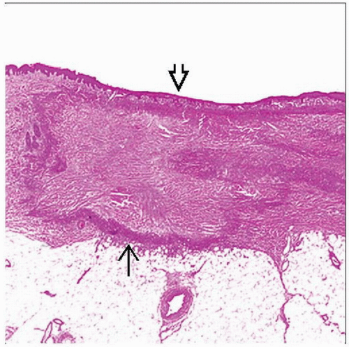
Small dermal/subcutaneous nodules with central necrosis
Epithelioid cells, spindled at periphery
Mildly atypical nuclei, eosinophilic cytoplasm
Rare multinucleated osteoclast-like cells
Rare myxoid change
Calcification in 20%, rare stromal bone formation
Occasional hemorrhage (“angiomatoid” or angiosarcoma-like variant)
Stay updated, free articles. Join our Telegram channel

Full access? Get Clinical Tree


