Figure 33-1. Heat sensor – nasal pit. (Photo courtesy of Ronny Stewart, MD.)
Snakes are easily identifiable reptiles, marked anatomically by the large number (greater than 120) of precloacal vertebral bodies. They also have a reduced (almost absent) left lung, a single ventricle (no ventricular septum), and no muscles in the ciliary body of the eye. Despite the absence of a ventricular septum, oxygenated and deoxygenated blood channels fairly separately though the heart.4 All snakes are strictly carnivores. The size of the snake dictates the size of the prey.
The pit vipers (the genus Crotalus, Sistrurus, and Agkistrodon) are morphologically distinguished by elliptical pupils, a triangular head, (Fig. 33-2) and retractable fangs. The fangs are tubular; injecting venom similar to a hypodermic needle. This is in contradistinction to the Elapids in which the fangs are fixed and grooved – wicking the venom into the victim. Venomous snakes have a single row of caudal plates distal to the anal plate, whereas harmless snakes have a double row of caudal plates. There are 32 species of rattlesnakes in the New World, ranging from southern Alberta to central Argentina.5–7 Rattlesnakes are only found in the Americas. All true rattlesnakes have a rattle (with the exception of an endangered species in Mexico – the Santa Catalina rattlesnake, which has only a vestigial rattle). The rattle is a series of interlocking segments. A new segment is added with each shedding of the skin, which may occur 2 to 3 times per year. There are two genus of rattlesnakes; Crotalus – the true rattlesnakes and Sistrurus – the pigmy rattlers. There are 14 species of Crotalus (Table 33-1) in North America. The largest species of rattlesnake in North America is the eastern diamondback (Crotalus adamanteus), which is slightly larger than its cousin the western diamondback (Crotalus atrox). There are eight members of the Agkistrodon genus – moccasins and copperheads (Table 33-2).

Figure 33-2. Triangular shape of pit viper head. (Photo courtesy of Ronny Stewart, MD.)
By far the most critical step in management of snakebite is identification of the offending species. The majority of snakebites are nonvenomous and require no treatment. Some bites, such as those of copperheads (Fig. 33-3) and pigmy rattlers (Table 33-3), are almost always self-limiting and require little more than observation. Others such as the diamondback (Fig. 33-4) and Mojave rattlers can require intense medical management, occasional surgical management, and even rarely cause death. Identification of the species by the patient is notoriously invalid; however, a detailed description of the offending snake should be taken. The only snake where color is useful in identifying is the coral snake–red and yellow kill a fellow, red and black venom lack (Fig. 33-5). Note that this is true only in North America, as there are venomous coral snakes in Central and South America that have red bands touching black bands. Other clues, such as geography and topography (proximity to water), can also at least help with identification of the genus. All snakebites should, at least initially, be considered an event with potential morbidity and possible mortality.
Table 33-1 Crotalus Species

Table 33-2 Agkistrodon Species

As snakes are cold-blooded animals, snakebites generally occur in the warm months, with a particular increase in incidence in the spring and fall. The majority of snakebites occur in young men, with up to 60% of bites occurring in victims that were deliberately provoking the snakes.
2 Venom is produced in Duvernoy glands on the dorsal sides of the head (corresponding to parotid glands) enhancing the triangular appearance. During envenomation the venom is forced through the fangs by the palantine muscles. If a fang is broken off, it will be replaced by the next in a row of “proto-fangs.” Any clothing may limit the amount and depth of envenomation. The purpose of the venom is to immobilize or kill the prey and begin digestion. Pit viper venoms are complex – with up to 50 different proteins and enzymes identified.8–10 Over 90% of the dry weight of venom is proteinaceous in nature. Although it is common to classify the venoms as neurotoxic or digestive in nature, all snakes have varying compositions of venoms. The Crotalids will have a higher percentage of digestive/enzymatic type proteins than the Elapids, which will contain a higher percentage of neurotoxins. Common enzymes are phospholipases, metalloproteases, collagenase, and hyaluronidase (Table 33-4). These peptides and polypeptides act by damaging vascular endothelium and other cellular membranes. Compliment is activated, fibrin is degraded, and platelets are activated. The increased permeability of the membranes leads to peripheral and pulmonary edema, hemorrhage, and hypotension. L-amino acid oxidase splits fibrinogen contributing to the induction of DIC. The Elapids in particular contain a higher percentage of proteins that block acetylcholine receptor sites. This can lead to dysphagia, slurred speech, seizures, coma, and death. Despite the very high toxicity of coral snake venom, only one fatality has been recorded in the United States in the past 50 years.

Figure 33-3. Copperhead. (Photo courtesy of Ronny Stewart, MD.)
Table 33-3 Sistrurus Species

3 When evaluating the snakebite victim, it is important to document both local and systemic signs and symptoms. Vital signs should be recorded, and repeated at intervals guided by the clinical response. Pain, edema, and erythema at the site are common. Secondary symptoms include muscle twitching, perioral paresthesias, and a metallic taste in the mouth. Marking lines at the site of documented discoloration (Fig. 33-6) can help the clinician follow the progression of the envenomation. Swelling in the affected digit or limb should be measured (circumference) and followed closely. The amount of local tissue destruction is an accurate marker of the degree of envenomation. It is probable that if there is no pain or erythema by the time that the patient arrives in the ED, no envenomation has occurred. Intravenous access should be obtained. Laboratory evaluation should include a CBC with platelet count, PT, PTT, CPK, fibrinogen level, creatinine, BUN, and urinalysis. Up to 30% of Crotalid envenomations will show an abnormality in the coagulation profile, although clinical bleeding is exceedingly rare.11 In Lovonas 2004 review of 400 copperhead bites from the Carolinas: no hypotension, no respiratory failure, and no deaths occurred.15 Eight patients did have laboratory abnormalities, but none developed bleeding complications. However, in severe envenomation by one of the larger rattlesnakes, diffuse capillary leakage can lead to pulmonary edema, hypotension, and shock.12 A consumptive coagulopathy may rapidly develop. Such patients may bleed spontaneously from any site. Acute renal failure may result from directs nephrotoxins as well as myoglobinuria. An electrocardiogram, or at least continuous cardiac monitoring should be considered. If there appears to be no evidence of envenomation, the patient may be observed in the emergency department for several hours and discharged home. With evidence of envenomation our recommendation is for admission to the hospital, preferably a monitored bed, for observation and possible intervention for at least 24 hours.

Figure 33-4. Western diamondback.

Figure 33-5. Coral snake.
Table 33-4 Composition of Venom


Figure 33-6. Demarcation of hemolysis after bite.
As noted above treatment is largely dictated by the species of snake and the initial clinical presentation. Although a number of series have noted the appropriate treatment of Agkistrodon (copperhead and moccasin) bite is observation only,13–15 extrapolation of this data to rattlesnakes could be misleading. In the rattlesnake bite victim with a moderate or severe envenomation, early use of antivenom is appropriate, and should be strongly considered (see below). Although there are a number of snakebite severity indices (grading systems) published, a modification of the simple system proposed by Dart in 1996 is probably the most useful.16 A grade 1 envenomation is limited to the immediate bite site, a grade 2 envenomation extends onto less than a full extremity and may have non–life-threatening symptoms such as nausea, vomiting, mild tachycardia, and mild hypotension. A grade 3 envenomation involves more than an extremity and includes systemic signs such as severe hypotension/tachycardia or blood dyscrasias or clinically significant clotting abnormalities. Although enzyme-linked immunosorbent assays (ELISAs) have been developed that can directly measure serum venom antigens as well as identify the offending snake, clinical applicability of these types of tests are limited.17
Initial first aid on the scene should involve immobilization and splinting. Although there are no data to confirm this will limit the spread of venom, it might be more comfortable. The application of ice or heat seems to have little effect on results. A number of historical treatments should specifically be avoided. Incision and suction, advocated for years, is no longer recommended.18 A surgical incision in the field by a nonsurgeon can possibly involve vessels, tendons, and nerves and increase morbidity, and only a small amount of venom can actually be extracted with suction. Limited cooling of the extremity may have some benefit, although a version of this – ligation cryotherapy – is associated with a high rate of amputation.19,20 Although placement of a constrictor band may be of benefit in Australia (where Elapids predominate) to prevent rapid dissemination of a deadly neurotoxin, it is probably of little benefit in North America where the offending species is almost certainly a Crotalid. The use of a compression bandage has been shown to slow the systemic absorption of venom.21,22 This may make it the treatment of choice in the field for a nonnecrotizing envenomation, such as produced by an Elapid. It may however increase local necrosis when used to treat the bite of a pit viper.
The wound should be cleansed and the location of the puncture sites documented. The distance between the two fang marks is related to the size of the snake. The tetanus immunization status of the patient should be reviewed and updated. Steroids have no benefit in humans although these are often used by veterinarians.23,24 Steroids do have a role in treating the infrequent acute allergic as well as delayed-onset serum sickness that may occur after the use of sheep-derived antivenom, as noted below.
The prophylactic use of antibiotics has been evaluated by several studies and does not seem to reduce infection rates.25–28 Infection after snakebite is surprisingly uncommon and while patients are often given an antibiotic, there is no good clinical evidence for their use. Although many patients will have erythema, edema, and tissue necrosis and will appear infected, these are usually sterile; culture-proven infection occurring after snakebite should be treated appropriately. Culture of organisms in the mouth of the rattlesnake include Pseudomonas, Enterobacteracie, Staphylococcus, and Clostridia.29
If the species is a coral snake or a nonindigenous Elapid, the administration of cholinergic agonists should occur at the first sign of symptoms. Calcium infusion may reduce the onset of seizures.
Certainly the most controversial decision point in management of snakebite is the administration of antivenom. Although hundreds of antivenoms are available worldwide for various species, for all practical purposes in North America we are only dealing with envenomation by Crotalids. Snakebite by coral snakes are uncommon, and while the venom is exceedingly toxic, the treatment is only supportive as antivenom is no longer available. Prior to 2000, the primary antivenom available in the United States for the bite of a Crotalid was a hyperimmune serum produced by envenomation of horses. Its use was associated with a high incidence of side effects: anaphylaxis, hypersensitivity, and delayed serum sickness.30 In 2000, CroFab (Crotalidae Polyvalent Immune Fab [Ovine]) was introduced obviating the use of now unavailable horse serum products.12,31
CroFab is truly an elegant drug. It is produced by immunizing sheep with the venom from the western diamondback rattlesnake (Crotalus atrox), the eastern diamondback rattlesnake (Crotalus adamanteus), the Mojave rattlesnake (Crotalus scutulatus) or the cottonmouth water moccasin (Agkistrodon). The monovalent immunoglobulin from the sheep is prepared by fractionation of the immunoglobulin from the serum, digesting it with papain, and then utilizing ion exchange and affinity chromatography to separate the Fab fragments specific to each species.32 The Fc portion of IgG is generally thought to be responsible for high rates of hypersensitivity and serum sickness seen with the horse serum product developed by Wyeth in 1954, and hence this ovine (sheep) version is generally considered safer. The four different monospecific antivenins are then mixed (Table 33-5). In a murine model there is good antigenic crossover in treating other North American Crotalid envenomations.33
Utilizing CroFab to treat Crotalid envenomation is somewhat controversial, primarily because of the cost/benefit ratio. The cost of this drug is a significant factor with wholesale prices up to $2,000 per vial. Certainly any patient with life-threatening symptoms should immediately be given 4 to 6 vials of the antivenom.34 Treatment is not based on the body mass of the patient – as the antivenom neutralizes the poison that is injected – this will vary with each envenomation. The lyophilized powder is reconstituted with 18 mL of normal saline, followed by dilution in 250 mL. Each dose is given over 1 hour. It is thought that venom is rapidly tissue bound, therefore the earlier the antivenom can be given, the more likely a favorable response. The packaging information states that treatment should begin within 6 hours of the bite. There is however at least one report of effective use of antivenom after significant delay.35 Thus is the conundrum – there are certainly snakebites that initially appear innocuous that later become life-threatening. This has lead physicians in the emergency room to over treat large numbers of patients who do not require therapy, and why it is so important that every attempt be made to identity the offending species. If one can ascertain that the snake is one of the less virulent types (Agkistrodon or Sistrurus species) the patient can almost certainly be treated without the use of antivenom.14,36 However, in the patient with a deteriorating clinical condition, treatment should always be given. Repeated dosing should be given until clinical stability is achieved; although this is highly unlikely after 30 to 40 vials. The patient should be monitored for recurrence of symptoms and retreated if necessary.37 CroFab has an estimated half-life of 12 to 23 hours.
Table 33-5 Components of Antivenom–CroFab Antibodies

Immediate adverse drug reactions will occur in 6% to 8% of patients.38,39 This may be related to the rate of infusion.40 Although anaphylaxis would occur in up to 50% of patients treated with the older equine antivenin, it is an uncommon occurrence with CroFab – maybe up to 14% of patients.41 It should be treated aggressively when occurring: The antivenin infusion should be stopped and a combination of antihistamines (diphenhydramine), epinephrine, and steroids administered, depending on the severity of the response. The airway should be assessed and secured if necessary. Appropriate volume resuscitation should be instituted. Steroid dosing ranges from a single 125 mg intravenous bolus of methylprednisolone to short-course prednisone pulse dosing (60 mg daily for 5 days).40
Serum sickness will occur in 13% to 16%39,41 of patients treated with ovine antivenom (CroFab), certainly less common than with the older Wyeth equine antivenom.30 Serum sickness is a type III hypersensitivity reaction in which soluble antigen–antibody complexes are deposited diffusely in the presence of antigen excess. Symptoms such as urticaria, itching, nephritis, and arthralgia can occur for weeks after the infusion. The treatment for serum sickness is a pulse of corticosteroids tapered over 7 to 14 days.
There is no question that the purified ovine antivenin (CroFab) is much safer than the older horse product.8,42 This has led to some indiscriminate usage in emergency departments across the country. It should be recognized that the vast majority of snakebite victims will recover uneventfully with only supportive treatment. Each patient must be individually clinically evaluated and limit antivenom use to those in whom the potential benefits outweighs the risk of side effect.
CroFab has been used extensively in children as young as 14 months.43–45 There are beginning to be reports of overuse of CroFab in children as well as adults.46 A special consideration should be use of antivenom in the pregnant patients. Certainly coagulopathy could be lethal to the mother and fetus. There have been limited reports of use of CroFab in pregnancy.47 Reports of fetal loss of up to 20% are noted in the worldwide literature with snakebites of a variety of species. This seems to be improved with the use of antivenom.47,48 Certainly the risk/benefit ratio must be carefully weighed, but the general recommendation is to treat pregnant women with CroFab when indicated.
In the rare patient that develops clinical bleeding; such as hemoptysis, intracranial hemorrhage, or gastrointestinal bleeding, correction of the coagulopathy should be undertaken with appropriate blood products. Antivenom should be given in these patients as initial therapy. Do not routinely treat thrombocytopenia or severe coagulopathy in the absence of bleeding. Coagulopathy can recur up to 2 weeks late.37

Figure 33-7. Hemorrhagic bullae.
The use of surgery in the management of snakebite has a limited role. Although historically snakebite has been treated with various surgical procedures,19,49 it should be in an unusual patient that more than simple debridement is needed. Crosscut and aspiration of the puncture sites was utilized for years, but this is no longer recommended. Several studies have shown the limitations of this practice. In dogs, up to one-half of the venom can be removed if incision and suction are begun within 3 minutes.17 In rabbits up to 37% of antivenom can be removed. However, in human studies only about 11% of the venom may be removed.50,51 It is rapidly bound to tissues and is difficult to be removed by suction. Laceration of vessels and nerves by well-meaning people at the scene is likely to increase morbidity as well as increase the risk of infection. Both partial and radical surgical excision of the involved bite area has likewise been attempted with no proven benefit.52–55 A common finding is a local cytolytic response (Fig. 33-7), that produces a hemolytic bullae and requires debridement (Figs. 33-8 and 33-9). This can be expected to heal by secondary intention with a satisfactory cosmetic response (Fig. 33-10).
Another use of CroFab is in the patient with impending compartment syndrome. The great majority of snakebites deposit venom in the subcutaneous tissues, not subfascial. It can sometimes be difficult to differentiate a swollen painful extremity from a true compartment syndrome. When in doubt compartment pressures should be measured.55,56 With rising pressures (greater than 30 to 40 mm), antivenom therapy should be initiated (or repeated). With failure of response to adequate dosing of antivenom, appropriate surgical fasciotomy can save extremities. Prophylactic fasciotomy has no role in treatment. Stewart and colleagues at the University of Texas Health Science Center in San Antonio have an extensive experience with rattlesnake bites.57 They have randomized animals to receive an intracompartmental injection of venom from the western diamondback rattlesnake. The animals received fasciotomy with debridement alone, antivenom alone, or antivenom with fasciotomy and debridement. Antivenom therapy prevented muscle loss and improved survival. Surgery alone did not. It was noted that muscle that had been debrided would have survived if treated with antivenom alone. Fasciotomy may increase the severity of local tissue loss.58,59 When fasciotomy is required because of rising compartment pressures that do not respond to antivenom, consideration for postoperative negative-pressure wound therapy should be given.

Figure 33-8. First debridement.

Figure 33-9. Second debridement.
Creative attempts at treating envenomation have even included the use of electrical current to neutralize the venom.60 The use of current from the generator of an outboard motor and an automotive battery has even been attempted. As innovative and entertaining as these efforts may be, there is no improved outcome.61 The TASER® or conductive electrical weapon (stun gun) is a small hand-held device that produces a very high voltage with a low current. It is used primarily by law enforcement agencies to temporarily immobilize attackers. A group of physicians practicing in Ecuador reported a series of 34 patients in letters to the editor of Lancet.62,63 They claimed an immediate improvement with no mortality. The snakebites were most likely Bothrops atrox – the Fer-de-lance – a pit viper. Unfortunately this reported treatment was not a controlled study. Several animal studies have since proven electrical shock therapy to be ineffective in snakebite.64–66
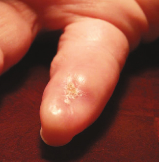
Figure 33-10. Healed wound.
Special Consideration of Elapid Envenomation
As noted there are only three species of Elapids in the United States – the eastern coral snake, the Texas coral snake, and the Arizona coral snake. The Eastern coral snake (Micrurus fulvius) is found in the southeastern United States up to Kentucky and North Carolina. The Texas coral snake (Micrurus tener) is found in west of the Mississippi across Arkansas, through Louisiana into eastern Texas. The Eastern and Texas coral snakes inhabit forests and shrubbery. The Arizona coral snake (Micruroides euryxanthus) is found in the desert climates of the southwestern United States into Mexico. Coral snakes are more active at night and hide under ground cover. Their behavior is elusive and they will generally try to flee when disturbed. This family of snakes has a common feature – bilateral upper maxillary fangs that are fixed into position and angled down and back. The fangs are grooved for instillation of the venom, which is predominately a neurotoxin. The snakes are oviparous and have round pupils. Their head is flat, with a smooth tapered tale and of course no rattles. They have characteristic coloration which is mimicked by several other nonvenomous species (such as the king snake and the scarlet king snake) in a biologic phenomenon referred to as Batesian mimicry.67 Although the North American coral snakes are relatively small (less than 5 ft), some Elapids from other continents are very large; the Black mamba is 14 to 15 ft and the King Cobra can be up to 18 ft and weigh 25 lb. Although nonnative elapids bites have a substantial risk for death, only one known death from a Coral snake has occurred in the United States in the last 50 years (Bonita Springs Florida 2006).68
The bite of an Elapid is likely to produce much less tissue injury than the bite of a Crotalid.69 There may be minimal pain. Envenomation may result in immediate neurotoxicity (within 15 to 30 minutes in the case of the mamba or Australian brown snake) or be delayed a bit (2 to 5 hours with coral snakes). There is no longer any antivenom available for North American coral snakes, but local zoos may be able to provide antivenom for more exotic Elapidae. First aid should involve a compression bandage over the wound. The poison generally spreads by lymphatics; therefore field compression to occlude lymph flow at the site of envenomation can slow toxicity. Symptoms will be related to the neurotoxicity – confusion, muscle spasm, nausea, vomiting, and dizziness. Blurred vision and difficulty with speech will occur. Finally, respiratory paralysis of the diaphragm occurs with resultant death. Cardiovascular and pulmonary support is essential to salvage patients that progress to this state.70 It should be noted that venom of an Elapid is incredibly toxic. The Taipan and Belcher sea snakes have an LD50 in mice of as little as 0.025 mg/kg of venom.71 Any suspected Elapid bite should be managed with the possibility of rapid demise of the patient. If the bite is of a nonnative Elapid, antivenom (if it can be found) should be administered promptly at the first sign of clinical demise. Death rates for untreated nonnative Elapids varies from 20% to 30% for cobras to nearly 100% for the mambas.72 Antivenom for exotic snakebites may be obtained from the local zoo or through their national organization – the Association of Zoos and Aquariums (https://www.aza.org) or through the Poison Control Center at 1-800-222-1222.

Figure 33-11. Gila monster.
Venomous Lizards
There are two venomous lizards in North America: the Gila monster (Heloderma suspectum) and the Mexican beaded lizard (Heloderma horridum). Both species have heavy bodies with large heads. The Gila monster is found in the southwestern United States, ranging primarily in Arizona, extending into southeastern California, southwestern Utah and New Mexico, and into northern Mexico. There are two subspecies: the banded Gila monster (Fig. 33-11) that is found in the northern portion of the habitat and the reticulated that exists in the southern portion of the range. It is the largest lizard in the United States with a length of up to 56 cm. The Gila monster has generated substantial lore in the southwestern United States, because of its characteristic appearance and apparent fetid breath. It lives below ground and spends most of its life in or near its burrow. It is primarily seen in the spring, in the morning hours during breeding season. It has powerful jaws and the bite is characterized by the tendency to hang on with the animal difficult to dislodge. The lizard has eight labial glands on either side of the lower jaw. The venom is secreted into the floor of the mouth where it is wicked into the victim via grooved lower canine teeth. Envenomation is limited and deaths are virtually unknown.73 The morbidity is usually secondary to local effects from the toxins present: serotonin, phospholipase A, amine oxidases, hyaluronidase, and proteases.74 These produce pain at the site that usually resolves in less than 24 hours. Hypotension, coagulopathy, and myocardial infarction have been reported.75 Nausea and vomiting can occur. Malaise may persist for several days. The patient should be admitted for observation and a low threshold for surgical exploration/debridement be given because the teeth often shed into the bite and can serve as a nidus for infection.76
The Mexican beaded lizard is found from Sonora to Guatemala and is represented by four subspecies. They are larger than the North American Gila monster; with a maximum size of up to 91 cm. They have similar venom as the Gila monster and bites should be managed in a supportive fashion.
Insect Bites and Stings
4 Although the bite of a venomous snake will certainly draw more attention in the emergency department, in fact insects kill many more Americans (greater than 100 deaths per year)77 than snakes (about 5 deaths per year). Although most of these deaths are from anaphylactic reactions, some are from overwhelming envenomation, particularly from those of the Africanized honey bee (the “killer” bee). Most of the treatment recommendations below are from expert opinion; few randomized trials have been performed.
Spiders
There are six genera of spiders in the United States that can produce painful bites, tissue necrosis, and rarely death. These fall into three groups – the brown recluse (Fig. 33-12), the black widow (Fig. 33-13), and the hobo spiders.78

Figure 33-12. Brown recluse.
Brown Recluse
There are 13 species of Loxosceles, of which 5 have been known to produce tissue necrosis. Probably the species producing the most pathology is the brown recluse (Loxosceles reclusa). Envenomation by the hobo spiders (Tegenaria agresis) produces a similar wound. The brown recluse is generally found in the southern states.79 Hobo spiders are found in the Pacific Northwest.80 Bites from these two species generally are initially painless and the victim may not recall being bit. Pain and erythema develop over the next 6 hours. The venom can produce a painful, necrotic, slow healing wound over the next 48 to 72 hours.79,81 Two to 7 days after the bite an eschar develops with surrounding soft tissue induration. The eschar can conceal an underlying tissue necrosis that can persist for months.79,82 These can require extensive debridement leading to skin grafting and even amputation.
The venom is both hemolytic and cytotoxic. The enzyme that is thought to produce the “desmonecrotic arachnidosis” is sphingomyelinase D. The venom also contains hyaluronidase, lipase, arachidonic acid, and prostaglandins.83,84 Systemic symptoms occur in less than 10% of victims, more commonly in children. Rarely hemolysis, hemodynamic instability, and death have occurred.85 Initial treatment should include analgesia, wound care, and tetanus prophylaxis. Antibiotics should be reserved for infection/cellulitis. Early debridement should be withheld. Consideration for oral dapsone therapy should be given. Dapsone is thought to work as an inhibitor of polymorphonuclear leukocyte chemotaxis. It has significant side effects and can produce hemolysis in G6PD-deficient patients. Its use is somewhat controversial. It has been shown to reduce the size of the skin lesion and lessen the amount of surgical debridement, but these are not prospective studies.86 A controlled study of dapsone, electroshock therapy and no treatment in guinea pigs showed some improvement with dapsone, but none with the electrical therapy.87 Hyperbaric oxygen has been studied in rabbits and pigs and failed to demonstrate significant improvement.88 A trial of hyperbaric oxygen, dapsone, and cyproheptadine in rabbits failed to show any improvement.89 Systemic steroids and antihistamines likewise have not been shown to help. Initial surgical excision of the lesion has no clear benefit.90 Delayed excision of the area of necrosis with skin grafting can be necessary. Recurrent necrosis in the same site can return even years later.
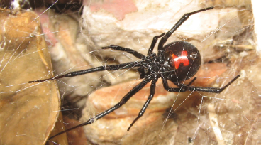
Figure 33-13. Black widow.
Black Widow Spiders
There are 31 species of the genus Lactrodectus, the Black Widow spider, of which 4 are found in North America. These are marked by a prominent red hourglass figure on the ventral aspect of the thorax. Although both the male and the female possess venom, only the bite of the female can penetrate human skin. The female is four times the size of the male. The female of the species is known for eating the male after mating, which may or may not occur. The bite may produce only minimal local symptoms, with only a small area of redness, or a “target” appearance. The venom contains alpha-latrotoxin, which is neurotoxic. This produces chest and abdominal pain, and the patient’s presentation may be confused as an acute surgical abdomen.91 Marked cramping and abdominal rigidity may be present. Fasciculations of facial muscles with ptosis and spasm may occur producing a particular facie. Hyperreflexia, paresthesia, cutaneous hyperesthesia, and nausea may also occur. Symptoms may occur within an hour and resolve over several days.92 The muscle spasm should initially be treated with benzodiazepines. Antivenin is available, and should be considered in the very young or ill patient. The dose is one ampule given in 50 mL of saline. The antivenom is equine and hypersensitivity testing should be done prior to giving the antivenom.93,94 A 1-mL test dose of horse serum is included with the antivenom for topical conjunctival testing. Calcium gluconate was often given in the past, but is no longer recommended. Mortality is exceedingly rare.
Hymenoptera
More than 100,000 species of Hymenoptera exist. This includes bees, wasp, hornets, and fire ants. The venom injected by these insects is at least as toxic as that of the rattlesnake, but the volume is much less. The venom is primarily a hemolysin and neurotoxin. Anaphylaxis is a common clinical problem produced by the envenomation. Approximately 0.4% of the human population is at risk of anaphylaxis from Hymenoptera stings.95 The anaphylaxis is modulated by preformed IgE that activates mast cells leading to massive histamine release. This leads to laryngeal edema, pulmonary edema, and cardiovascular collapse. This must be treated aggressively with intravenous diphenhydramine and epinephrine as well as with management of the airway and supplemental oxygen. Most stings are mild, however, resulting in only dermal reactions – pain, erythema, and edema. The stinger should be removed and ice applied to the site. Application of meat tenderizer might be of some benefit. Patients with a history of insect sting anaphylaxis should be given a prescription for an epinephrine pen and be considered for venom immunotherapy. This is almost 100% successful in desensitization, but can take up to 3 years.
Killer Bees
Africanized honeybees (killer bees) are hybrids of the African honeybee and the European honeybee, the one commonly found in the United States. The Africanized honeybee was originally brought from Tanzania to Brazil in 1957, with the hope of better honey production. The bees have spread up through Central America, Mexico, and into Texas. They are continuing to advance at about 100 miles per year.96,97 Africanized honeybees are indistinguishable from the European honeybee, so DNA (usually mitochondrial DNA) analysis is necessary to differentiate the subspecies.98 They are termed “killer bees” because of increased aggressive traits. They show greater defensiveness of the hive, they have a bigger “alarm” area, and they deploy in greater numbers and will pursue perceived threats over much greater distances. Although their venom is of the same potency as the European bees, the number of stings received will usually be much greater. With more than 50 stings, nausea, vomiting, shock, hemolysis, rhabdomyolysis, coagulopathy, coma, and death may occur. Usually most patients will just complain of pain, but delayed toxic reactions – 6 to 48 hours – may also occur. It is recommended that any patient presenting with more than 50 stings be hospitalized and observed.99 Laboratory evaluation with coagulation studies, platelet count, and liver function studies should be obtained.
Fire Ants
Fire ants are wingless members of the Hymenoptera order. They were brought to the United States from South America around 1918.100 The red imported fire ants appear to have come over on a ship through Mobile, Alabama. Shipments of infested nursery stock and other agricultural products, natural mating flights, and floating on flood waters have contributed to their spread. Two species of imported fire ant now infect large areas of the Gulf Coast states.101 Fire ants aggressively attack any perceived danger to themselves or their mound.102 Each ant can produce multiple stings. The venom is a dialkylpiperidine that induces the release of histamine from increased mast cell reactivity. The fire ant sting produces a sterile pustule, which does not usually require antibiotic therapy. Infectious complications can occur at the site that may be more common in diabetic or immunocompromised individuals. Fire ant venom shares at least four antigenic proteins with bees and wasps. More than 80 fatalities have been reported from fire ant anaphylaxis, notably in Australia and southern United States. The treatment is the same as with bee/wasp sting anaphylaxis.77
Scorpions
More than 1,500 species of scorpions can be found worldwide. Only about 20 or 25 are considered dangerous. They are in the class Arachnidia; close relatives of ticks, mites, and spiders. Their characteristic shape makes them readily identifiable. Scorpions prefer dry habitats but may be found throughout the southern United States. Scorpions are nocturnal, hiding during the day and becoming active at night. This behavior helps them manage their temperature and water balance, important for survival in their dry habitats. The scorpion’s body has a 5-segmented tail that can be arched over the back and becomes more slender toward the end. On the end of the tail is the bulb-like poison gland/stinger. Between the last pair of legs are comb like structures called the pectines – sensory organs used to sense surface textures and detect prey. The venom toxicity varies greatly depending on the species, season, and age of the scorpion. Stings from dangerous species may cause paralysis, severe convulsions, cardiac irregularities, breathing difficulties, and death. In the United States most scorpion bites are not life-threatening and only result in local effects. In parts of Brazil, Mexico, North Africa, and Israel, however, the sting may be lethal. In Brazil the mortality rate is as high as 12% for adults and 60% in children.103 In India, of 34 children admitted to a hospital for a scorpion sting, 14 had hypertension, 9 had acute pulmonary edema, 5 had a myocardial infarction, and 4 died.104 Antivenins are typically available in area where dangerous species are found, either from a local zoo or state or university agricultural extension departments. One species in the North America is particularly venomous – the Arizona bark scorpion (Centuroides sculpturatus) (Fig. 33-14). The sting produces a dramatic neuromotor syndrome and respiratory insufficiency. It has produced several deaths in the United States and hundreds in Mexico. An antivenin is available in Mexico, but not in the United States.

Figure 33-14. Arizona bark scorpion.
Centipedes and Millipedes
There are approximately 3,000 species of centipedes in the world. They are arthropods with a single set of legs on each body segment. The front pair of legs are modified to deliver venom. One of the largest members of this class is the giant desert centipede (Scolopendra gigantea). It can grow to 26 cm in length. Significant morbidity, including EKG changes and rhabdomyolysis, can occur from centipede bites, but death is exceedingly rare. The most common symptoms include erythema, swelling, and pain.105 Treatment is largely symptomatic, with reports of both ice and heat improving symptoms.106 There are approximately 7,000 species of millipedes. They have 2 pairs of legs on each body segment. Millipedes do not have a mechanism to inject a venom, but instead secrete an irritating liquid from pores covering their body.107 This can produce a superficial burn that is self-limiting. Only symptomatic therapy is indicated.
Asp Caterpillars
The asp is a venomous caterpillar found in the southern United States. It is the larval stage of the Southern Flannel Moth (Megalopyge opercularis) (Fig. 33-15). It has the appearance of soft furry ball, encouraging the unwary to touch it. The long hairs of the asp conceal shorter spikes that discharge venom. Typically an intense throbbing and pain occurs almost immediately. Erythematous spots occur at the site of envenomation. Symptoms include headache, nausea, vomiting, and the development of lymphadenopathy. Chest pain occurs occasionally. Rarely shock and respiratory distress have been reported. Death is exceedingly rare and is almost certainly because of a hypersensitivity reaction.108–110 A similar caterpillar found in South America – Lonomia – has a death rate of 1.7%. The initial treatment should be to apply adhesive tape and pull it off to remove the spines. Ice packs and antihistamines may provide symptomatic relief. Intravenous calcium and steroids have been suggested as a treatment regimen.111

Figure 33-15. Asp caterpillar.
Hypothermia
5 Humans will always attempt to maintain a constant body temperature despite changes in environmental temperature. Normal body temperature is 37°C sublingually, 38°C in the rectum, 32°C at the skin, and 38.5°C deep in the liver, with normal daily (circadian) variation of about 0.5°C to 1°C; we tolerate poorly even minor deviations from these norms.112 Although humans have a remarkable capacity to dissipate heat by evaporating body water, our tropical evolutionary heritage has provided us with far less ability to cope with cold conditions. As a result, hypothermia can occur in a variety of clinical settings and from a number of causes (Table 33-6).
Table 33-6 Clinical Definitions of Hypothermia and Examples of Settings in Which They Occur


Figure 33-16. Number of hypothermia-related deaths by year in the United States, 1979–2002. (After Hypothermia-related deaths—United States, 2003–2004. MMWR Morb Mortal Wkly Rep 2005;54:173–175.)
Hypothermia is considered to be present in humans if the core temperature drops below 35°C (95°F). Hypothermia is usually classified by temperature zones: mild (32°C to 35°C); moderate (28°C to 32°C); or severe (<28°C). Primary accidental hypothermia is defined as a decrease in core temperature that occurs as a result of overwhelming environmental cold stress, such as recreational misadventures that lead to cold-water immersion or prolonged environmental exposure. Secondary accidental hypothermia occurs in patients with abnormal heat production or thermoregulation, who become cold despite only mild cold stress. The most significant risk factors are advanced age, mental impairment, and substance abuse (alcohol, primarily), although hypothyroidism, hypoadrenalism, trauma, and hypoglycemia are other risk factors.113,114 Chronic hypothermia develops in patients with impaired heat generation (i.e., the elderly and infirm) who live in unheated apartments, are under continual cold stress, and after a time are found to have a chronically low temperature as if they have autoregulated to a new, lower-set core temperature.
A multicenter review of 428 cases of accidental hypothermia reported an overall mortality rate of 17%,115 although other reports document mortality rates as high as 80%, primarily a result of infection and underlying illness. Treatment of accidental hypothermia remains highly variable. A report from an academic medical center in Amsterdam highlights this, as they reported that over an 8-year period (2000 to 2008) a total of 84 patients with accidental hypothermia were treated with 14 different rewarming techniques, with an overall mortality of 28.6%.116 It is estimated that each year about 1,500 patients in the United States have hypothermia listed on their death certificate, although the exact incidence of primary and secondary hypothermia and the associated morbidity and mortality remain uncharacterized.114 From 1979 to 2002, the CDC reported that a total of 16,555 deaths in the United States, an average of 689 per year (range 417 to 1,021), were attributed to exposure to excessive natural cold (Fig. 33-16).113 From 1999 to 2002, a total of 4,607 death certificates in the United States had hypothermia-related diagnoses listed as the underlying cause of death for an annual incidence of 4 per 1,000,000 population.113 Most reported hypothermia-related deaths (67%) occurred in males (Fig. 33-17), but the overall death rate is the same for both males and females. Notable is that deaths occur in all states, even those that generally are considered to have warm climates. Hypothermia-related deaths are reported by states with characteristically milder climates that experience rapid temperature changes and by western states that have high elevations and experience considerable changes from daytime to nighttime temperatures. States with the greatest overall death rates caused by hypothermia are Alaska, New Mexico, Wyoming, and Montana (Fig. 33-18).

Figure 33-17. Number and rate (per 100,000 population) of hypothermia-related deaths, by age group and gender, in the United States, 1979–2002. (After Hypothermia-related deaths United States, 2003–2004. MMWR Morb Mortal Wkly Rep 2005;54:173–175.)
Stay updated, free articles. Join our Telegram channel

Full access? Get Clinical Tree


