Embryonal Rhabdomyosarcoma

 1% of all rhabdomyosarcomas occurring in children


image Most common malignant neoplasm of extrahepatic biliary tree in children


• Occasionally seen in adults, usually in gallbladder

• Long-term survival can be achieved with modern multimodality therapy
image Estimated 5-year survival rate: 66%


Macroscopic


• More common in biliary tree than gallbladder
image Common bile duct is most frequent location

image Also reported in hepatic ducts, cystic duct, and ampulla of Vater

• Polypoid or grape-like (botryoid) gelatinous masses in lumen of bile duct or gallbladder

• May extend into liver or adhere to adjacent organs such as duodenum, stomach, and pancreas


Microscopic


• Tumor cells densely packed beneath single layer of biliary epithelium to form characteristic “cambium layer”
• Tumor cells typically have small round or ovoid hyperchromatic nuclei and variable amounts of eosinophilic cytoplasm

• Spindled cells may be present and can be prominent

• Varying numbers of rhabdomyoblasts

image Large round or elongated cells with abundant eosinophilic granular or fibrillar cytoplasm

image Elongated cells are referred to as strap cells

image May show cytoplasmic cross striations


Ancillary Tests


• Myogenic differentiation shown by positive immunostains for myogenin, MYOD1, desmin, muscle specific actin

image
Mass in Common Hepatic Duct
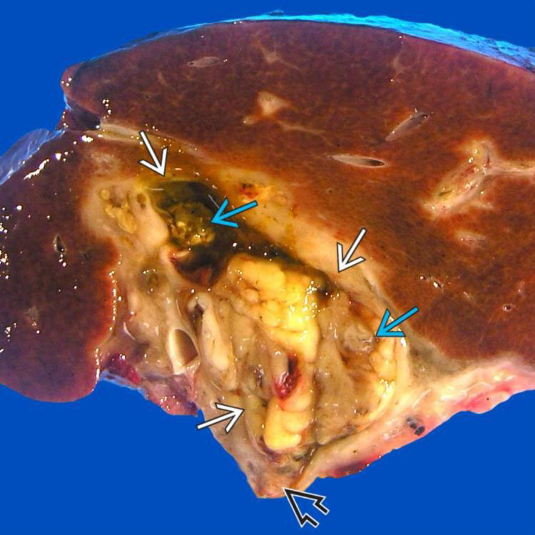
Liver of a 2-year-old girl shows a large embryonal rhabdomyosarcoma (RMS) in the porta hepatis. Mass image obstructs common hepatic duct image and extends to hepatic duct resection margin image. It shows a variegated cut surface due to necrosis caused by neoadjuvant chemoradiation therapy.


image
Cambium Layer
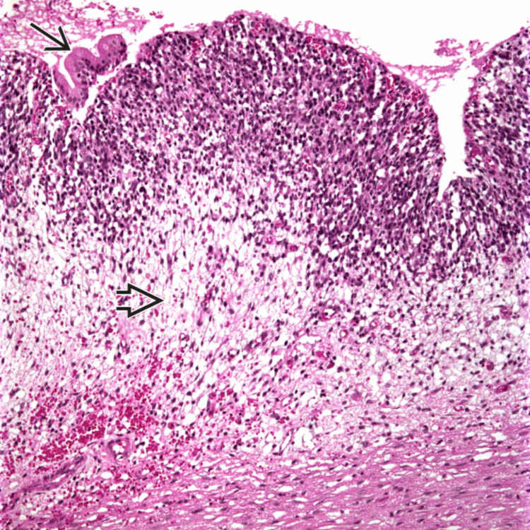
Characteristic cambium layer consists of densely packed tumor cells beneath biliary epithelium image, which is largely denuded. Deeper portion of tumor is hypocellular with loose myxoid stroma image .

image
Small Round Tumor Cells
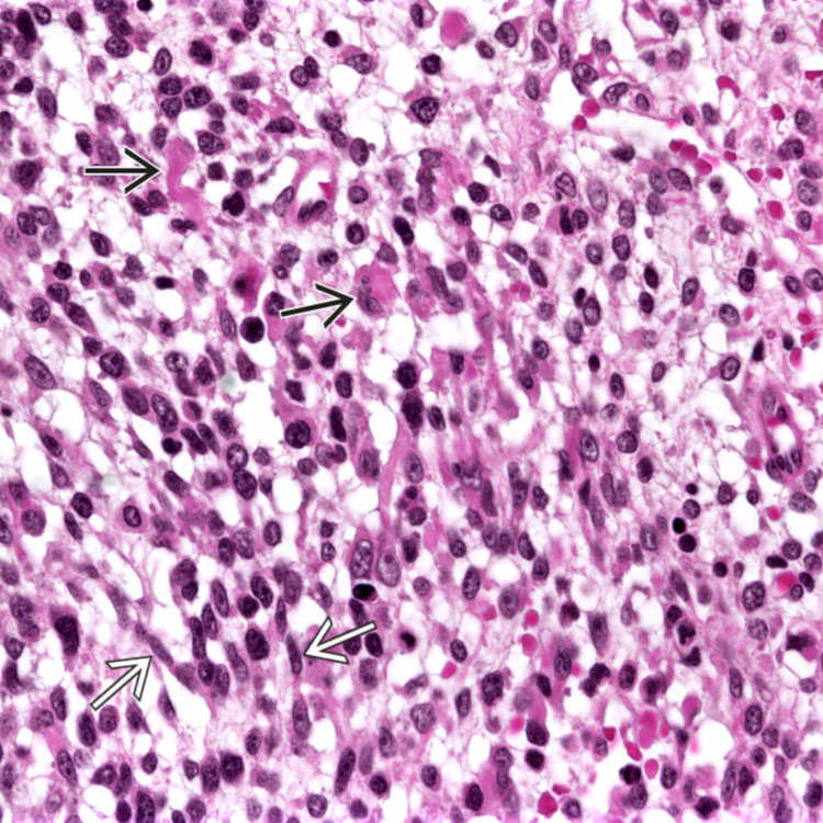
Tumor cells in embryonal RMS are typically small, round or ovoid, and hyperchromatic. Spindled cells can be present image. Occasional rhabdomyoblasts  are seen, which have abundant eosinophilic cytoplasm image .

image
Strap Cells
This case of embryonal RMS shows a focal area with multiple elongated strap cells with abundant eosinophilic cytoplasm image
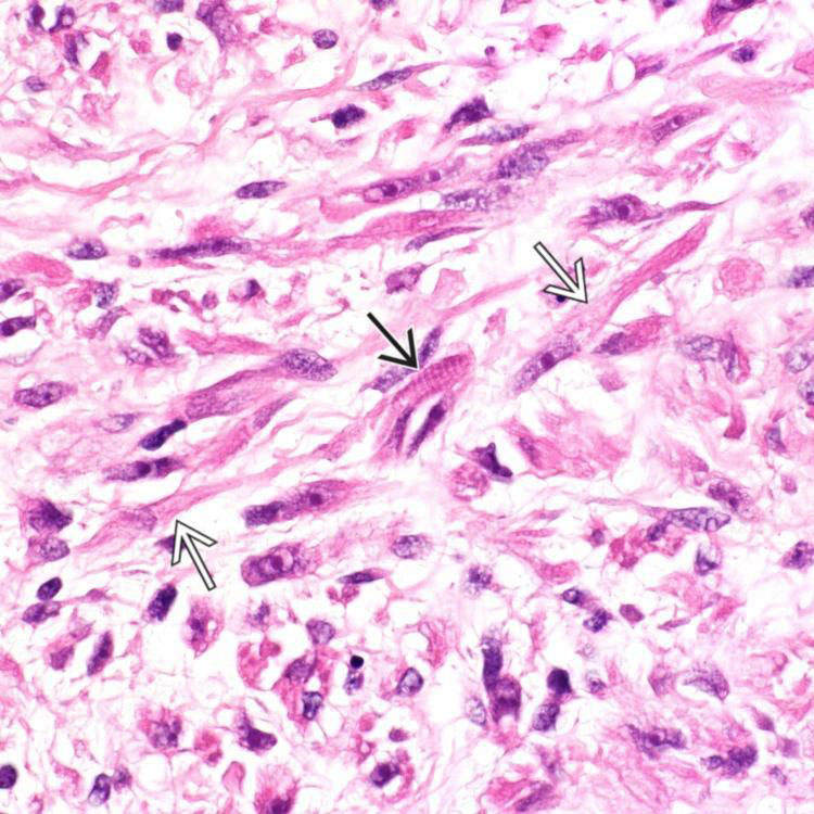
. Cytoplasmic cross striations are noted in one cell image .


TERMINOLOGY


Abbreviations


• Rhabdomyosarcoma (RMS)


Definitions


• Primary RMS, embryonal type, arising in biliary tree and gallbladder


CLINICAL ISSUES


Epidemiology


• Age
image Most frequently described in children, in extrahepatic biliary tree

Only gold members can continue reading. Log In or Register to continue

Stay updated, free articles. Join our Telegram channel

Apr 20, 2017 | Posted by in PATHOLOGY & LABORATORY MEDICINE | Comments Off on Embryonal Rhabdomyosarcoma

Full access? Get Clinical Tree

Get Clinical Tree app for offline access